Mostrando entradas con la etiqueta unit 4. Mostrar todas las entradas

martes, 17 de mayo de 2011

Skeleton vocabulary

I'm sure some of you were missing the post about the vocabulary:


  • Bone marrow Soft connective tissue found inside many bones; site of blood cell formation.

  • Cartilage Smooth covering found at the end of bones; made of tough collagen protein fibers; creates smooth surfaces for the easy movement of bones against each other.

  • Compact bone The dense, hard outer layer of a bone.

  • Joint Point at which two or more bones meet.

  • Ligaments Fibrous tissue that connects bones to other bones; made of tough collagen fibers.

  • Movable joint Most mobile type of joint; the most common type of joint in the body.

  • Skeletal system Body system that is made up of bones, cartilage, and ligaments.

  • Skeleton Sturdy scaffolding of bones and cartilage that is found inside vertebrates.

  • Spongy bone Lighter and less dense than compact bone; found toward the center of the bone.

  • Sprain A ligament injury; usually caused by the sudden overstretching of a joint which causes tearing.

martes, 10 de mayo de 2011

Joints and How they move

A joint is a point at which two or more bones meet. There are three types of joints in the body:
Fixed joints is a joint between two bones that doesnt move . Many of the joints in your skull are fixed joints. The skull plates don't move together or against each other, but they are connected or fused
Partly movable joints allow only a little movement..Your backbone has partly movable joints between the vertebrae. They are connected to each other by pads of cartilageand ligaments. They can only move a small amount
Movable joints allow movement and provide mechanical support for the body. Movable joints are the most common type of joint in your body. Your fingers, toes, hips, elbows, and knees all have movable joints. The surfaces of bones at movable joints are covered with a smooth layer of cartilage. The space between the bones in a movable joint is filled with a liquid called synovial fluid. The fluid lubricates and cushions the bones when they move at the joint. Ligaments help provide their stability and muscles contract to produce movement.
The knee joint is the largest joint in the body, consisting of 4 bones- the femur (thigh bone), the tibia (shin bone), fibula (outer shin bone) and patella (kneecap)- and an extensive network of ligaments and muscles..
There is a joint capsule that is a thick ligamentous structure that surrounds the entire knee. Inside this capsule is a specialized membrane known as the synovial membrane which provides nourishment to all the surrounding structures. The capsule itself is strengthened by the surrounding ligaments.
The stability of the knee owes greatly to the presence of its ligaments. Each has a particular function in helping to maintain optimal knee stability in a variety of different positions. There are two Collateral Ligaments ( inner and outer side) and two Cruciate ligaments.
Each knee joint has two crescent-shaped cartilage menisci. These lie on the medial (inner) and lateral (outer) edges of the upper surface of the tibia bone. They are essential components, acting as shock absorbers for the knee as well as allowing for correct weight distribution between the tibia and the femur.

domingo, 8 de mayo de 2011

Appendicular Skeleton


Appendicular Skeleton The appendicular skeleton consists of the bones of>the upper and lower extremities.
UPPER EXTREMITY
The upper extremity consists of the bones of the shoulder (pectorla girdle), the arm, the forearm, the wrist, and the hand.

  • Clavicle.—The clavicle (commonly called the collar bone) The clavicle and the scapula forms the pectoral girdle
  • Scapula.—The scapula is a triangular bone that lies in the upper part of the back
  • Humerus.—The humerus is the longest bone of the upper extremity and is often called the arm bone It articulates with the pectoral girdle to form the shoulder joint, and with the bones of the forearm to form the elbow
  • Radius and Ulna.—When the arm is in the anatomical position with the palm turned forward, the radius is on the lateral (thumb) side and the ulna is on the medial (little finger) side of the forearm
  • Carpal.—There are eight carpal bones, arranged in two rows, forming the wrist. Metacarpal.—The metacarpal bones are in the palm
  • Phalanges.—The small bones of the fingers are called phalanges, and each one of these bones is called a phalanx. Each finger has three phalanges, except the thumb (which has two)
LOWER EXTREMITY.—The lower extremity includes the bones of the hip (pelvic girdle), thigh, leg, ankle, and foot
  • Pelvic bones.— Commonly known as the hip, is a large, irregularly shaped bone composed of three parts: the ilium, ischium, and pubis
  • Femur.—The femur, or thigh bone, is the longest bone in the body (fig. 1-24).
  • Patella.—The patella is a small oval-shaped bone overlying the knee joint. It is enclosed within the tendon of the quadriceps muscle of the thigh.
  • Tibia.—The tibia, or shin bone, is the larger of the two leg bones and lies at the medial side.
  • Fibula.—The fibula, the smaller of the two leg bones, is located on the lateral side of the leg, parallel to the tibia.
  • Tarsus.—The tarsus, or ankle, is formed by seven tarsal bones
  • Metatarsus.—The sole and instep of the foot is called the metatarsus and is made up of five metatarsal bones
  • The phalanges are the bones of the toes and are similar in number, structure, and arrangement to the bones of the fingers

Axial Skeleton

DIVISIONS OF SKELETON
The human skeleton can be divided into two main parts; the first part is the axial skeleton and second part is the appendicular skeleton.
Axial Skeleton:
The axial skeleton consists of the skull, thevertebral column, and the thorax.

SKULL.— The skull is the bony structure in the head. It supports the structures of the face and forms a cavity for the brain.The skull is composed of two parts: the cranium and the mandible

Cranial Bones.—The cranium is formed by eight major bones, most of which are in pairs.
The frontal bone forms the forehead and the nasal cavity.
The parietal bones form the roof of the skull.The temporal bones help form the sides and base of the skull and also house the auditory and hearing organs.
The occipital bone forms part of the base and back of the skull, and contains a large hole called the foramen magnum
The sphenoid bone is situated on the base of the skull
The ethmoid bone is located in front of the sphenoid
Facial Bones.— : 13 immovable bones and a movable lower jawbone
The maxillary bones form the upper jaw, the anterior roof of the mouth, the floors of the orbits, and the sides and floor of the nasal cavity
The lower jawbone is called the mandible

VERTEBRAL (SPINAL) COLUMN
The vertebral column consists of 24 movable or true vertebrae; the sacrum; and the coccyx or tail bone. The bodies of the vertebrae are separated from each other by intervertebral discs

The spinal column is divided into five regions in the following order:
Cervical (neck), seven vertebrae
Thoracic (chest) twelve vertebrae that articulate with the ribs
Lumbar five lumbar vertebrae located in the small of the back, these vertebrae are the larger and stronger segments of the vertebral column
Sacrum is the triangular bone immediately below the lumbar vertebrae. It is composed of five fused vertebrae The sacrum is connected on each side with the hip bone and with the coccyx to form the posterior wall of the pelvis.
Coccyx is the tailbone. It have four fused vertebrae
THORAX.
The skeleton of the thorax includes 12 ribs on each side articulates posteriorly with the thoracic vertebrae and the sternum in the front upper half of the chest wall.
ribs 1 to 7 are connected to the sternum' by their costal cartilages and are called true ribs
ribs 8 to 10 are termed false ribs. They join the costal cartilage immediately above.by means of their costal cartilages.
ribs 11 and 12, which are free, are known as floating ribs.

Structure of Bones

Bones are rigid organs that are made up of two main types of bone tissue:
Compact bone makes up the dense outer layer of bones.
Spongy bone is lighter and less dense than compact bone, and is found toward the center of the bone. The tough, shiny, white membrane that covers all surfaces of bones is called the periosteum
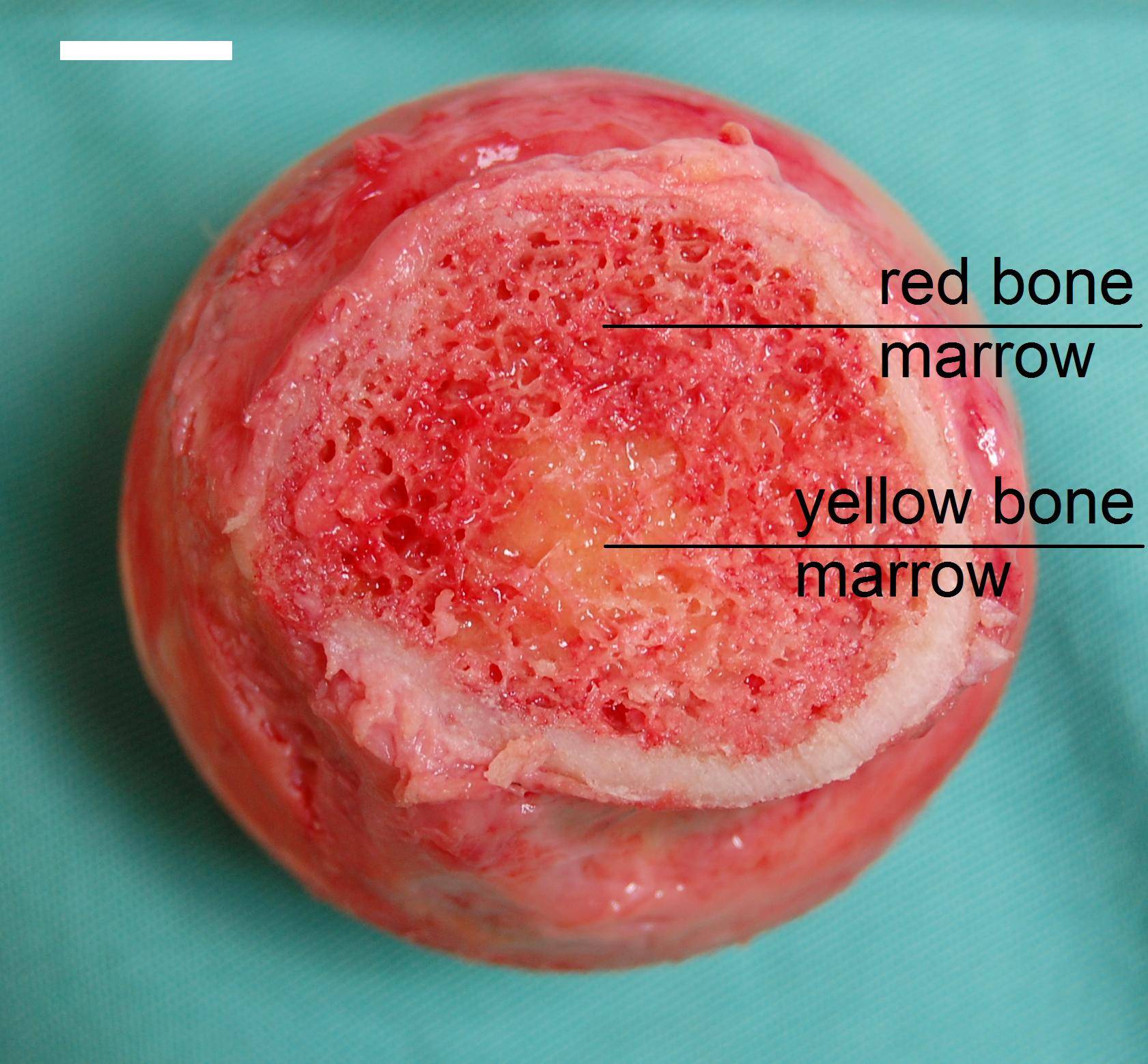
Many bones also contain in their innermost part a soft connective tissue called bone marrow. There are two types of bone marrow: red marrow and yellow marrow. Red marrow makes blood cells. Yellow marrow consists of fat cells although it will have a different role to that of normal fat cells
The bones of newborn babies contain only red marrow. As children get older, thei red marrow is replaced by yellow marrow. In adults, red marrow is found mostly in the bones of the skull, the ribs, and pelvic bones.

Classification of bones
There are four main types of bones in the human body. They can be long, short, flat, or irregular. Identifying a bone as long, short, flat, or irregular is based on the shape of the bone not the size of the bone.
Long bone are longer than wide . They consist of a long shaft (diaphysis) with two bulky ends or extremities(epiphyses). The structure of a long bone is shown in Figure. They are primarily compact bone but may have a large amount of spongy bone at the ends or extremities. Long bones include femur, tibia, and fibula of the leg, the humerus, radius, and ulna of the arm and the phalanges of the fingers and toes.
Short bones are roughly cube shaped with vertical and horizontal dimensions approximately equal. They consist primarily of spongy bone, which is covered by a thin layer of compact bone. Short bones include the bones of the wrist and ankle.
Flat bones are thin, flattened, and usually curved. Most of the bones of the cranium are flat bones.
Irregular bones are not in any of the above three categories. They are primarily spongy bone that is covered with a thin layer of compact bone. The vertebrae and some of the bones in the skull are irregular bones.
In this video you can watch the different types of bones:

Skeleton

Humans are vertebrates, which are animals that have a backbone. The sturdy scaffolding of bones and cartilage that is found inside vertebrates is called a skeleton. The adult human skeleton has about 206 bones, some of which are named in Figure.
The skeletal system is made up of bones, cartilage, and ligaments.
Bones are not only the hard mineral part as the remains that you might see in a museum. Living bones are full of life. They contain nerves, blood vessels and many different types of tissues
Cartilage is found at the end of bones and is made of tough protein fibers called collagen. Cartilage creates smooth surfaces for the movement of bones that are next to each other, like the bones of the knee.
Ligaments are made of tough protein fibers and connect bones to each other.
Functions of Bones
Your skeletal system gives shape and form to your body, but it is also important in other homeostatic functions. The main functions of the skeletal system are:
  • Support The skeleton supports the body against the pull of gravity. The large bones of the lower limbs support the trunk when standing.
  • Protection The skeleton provides a framework that supports and protects the soft organs of the body. For example, the skull surrounds the brain to protect it from injury. The bones of the rib cage help protect the heart and lungs.
  • Movement Bones work together with muscles as simple mechanical lever systems to move the body.
  • Making Blood Cells Blood cells are made mostly inside certain types of bones.
  • Storage Bones store calcium. They contain more calcium than any other organ does. Calcium is released by the bones when blood levels of calcium drop too low. The mineral phosphorus is also stored in bones.
As in other units in this link Kidshealth you can read and listen information about this subject, and finally a video with a reviw of all about skeleton.

Hormonal Regulation

Hormones are important to homeostatic regulation because they regulate many cell activities. We are going to see two different regulation:
  • Homeostatic feedback control mechanisms
  • Hormone antagonists.
Feedback control mechanism is a signaling system in which a product or effect of the system controls an earlier part of the system, either by shutting the process down (Negative feedback) or speeding it up (positive feed back). Most feedback mechanisms of the body are negative, only a few are positive.
Negative Feedback:
Negative feedback is a reaction in which the system responds in such a way as to reverse the direction of change. Since this tends to keep things constant, it allows for a process to return from a state of imbalance back to a homeostatic equilibrium. The thermostat is a good example to understand the negative feedback:
An example of negative feedback in the body is the control of blood-glucose concentrations by insulin. A higher amount of glucose in the blood signals the pancreas to release insulin into the blood. Then, the blood glucose concentration decreases and this lower concentration causes a decrease in the secretion of insulin by the pancreas.
Another example of negative feedback is the Regulation of pituitary glands and tropic hormans as you can see in the figure:
Positive Feedback
Positive feedback is a reaction in which the system responds in such a way as to speed up the direction of change. Positive feedback mechanisms are not as common as negative one.
Hormone antagonists
Many hormones work with hormone antagonists to control the concentrations of substances in the body. The hormones have opposite actions on the body and so are called antagonistic.
Insulin and glucagon make up an antagonistic hormone pair. The action of insulin is opposite that of glucagon. When your blood glucose concentration rises sharply after you eat food that contains simple carbohydrates, the increase in blood glucose level stimulates the pancreas to release insulin into blood. In response to signals by insulin most body cells take up glucose, which removes it from the blood, and the blood glucose concentration returns to the set point.
Later, when your blood glucose concentration has dropped below the set point, the decrease in glusose stimulates the pancreas to release glucagon. Glucagon causes the release of glucose from liver cells, which increases your blood-glucose concentration. This antagonistic relationship between the two hormones helps to maintain the narrow range of blood glucose concentration.

jueves, 5 de mayo de 2011

Adrenal glands

Adrenal Glands
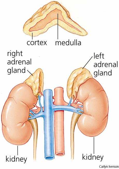
An adrenal gland is located above each of the kidneys, as shown in Figure. Each adrenal gland is separated into two structures, the adrenal medulla, which is the center of the gland, and the adrenal cortex, which is the outer layer. The medulla and the cortex work as two separate endocrine glands.
Adrenal medulla
It is the core of the adrenal gland, and is surrounded by the adrenal cortex. Secretion of hormones from the medulla is controlled by the sympathetic nervous system. It produce the hormones adrenaline (epinephrine) mainly.This hormone is part of the fight-or-flight response initiated by the sympathetic nervous system. It plays central role in short-term response to stress, increases heart rate and supply of blood with oxygen and glucose to the brain and muscles, while suppressing other non-emergency bodily processes, such as digestion.

Adrenal cortex
Is is the site of steroid hormone synthesis. In contrast to the medulla that is controlled directly by the nervous system, the cortex is regulated by hormones secreted by the pituitary gland. In adrenal cortex are produced:
Cortisol, often called the "stress hormone" as it is involved in the response to stress, and is involved in restoring homeostasis after a stressful event. Cortisol increases blood pressure, blood sugar levels and has an immunosuppressive action.
Aldosterone regulates balance of salt and water in the body. It stimulate sodium reabsorption in the kidneys. This increases blood volume and, therefore, increases blood pressure

Gonads
The ovaries of females and the testes of males are the gamete producing organs, or gonads. Ovaries in females are homologous to testes in males. In addition to producing gametes, an exocrine action, the gonads are endocrine glands that produce steroid sex hormones. Sex hormones secretion is controled by the gonadotropines produced in pituitary gland.
Testes
The sex hormones produced in testes are called androgens. The main androgen produced by the testes is testosterone. Testosterone is responsible for development of the male sex organs and secondary sex characteristics of males such as facial hair and deepening of the voice .
Ovaries
In females the ovaries produce two hormones: estrogen and progesterone. Estrogen causes the release of an egg from the ovaries and progesterone prepares the uterus for a possible implantation by a fertilized egg. Both hormans take part in control the menstrual cycle.
The placenta is an endocrine gland of pregnancy because it secretes estrogens and progesterone which are important for maintaining a pregnancy.

Other Endocrine Glands I

Thyroid Glands
The thyroid is one of the largest endocrine glands in the body. This butterfly-shaped gland is found in the neck, wrapped around the trachea, as shown in Figure. The thyroid is controlled by the pituitary.
The main thyroid hormone is thyroxine (T4). It controls the pace of all of the processes in the body. This pace is related to your metabolism. If there is too much thyroid hormone, every function of the body tends to speed up. As thyroxine controls how quickly the body burns calories, the thyroid gland regulates the body temperature by secreting more or less hormones.
The element iodine is very important for making thyroxine. If a person’s diet does not have enough iodine, their thyroid cannot work properly and the person develops an iodine deficiency disease called goiter. The addition of small amounts of iodine to table salt, has helped reduce the occurrence of iodine-deficiency in developed countries
As a result, problems with the under secretion (Hypothyroidism) or over secretion (Hyperthyroidism) of thyroid hormones affect many body systems.
Pancreas
The pancreas is both an exocrine gland as it secretes pancreatic juice containing digestive enzymes, and an endocrine gland as it produces several important hormones. It is located just below and behind the stomach, as shown in Figure. The endocrine cells of the pancreas are grouped together in areas called islets of Langerhans, shown in Figure. The islets produce the hormones insulin and glucagon. Insulin and glucagon are both involved in controlling blood glucose levels. Insulin causes excess blood glucose to be taken up by liver and muscle cells, where it is stored as glycogen, a polysaccharide. Glucagon stimulates liver cells to break down stores of glycogen into glucose which is then released into the blood.
So:

  • Insulin reduces blood glucose concentration

  • Glucagon raises blood glucose concentration

Hypothalamus and pituitary gland

The hypothalamus is a portion of the brain located in its lower central part. The hypothalamus is the primary link between the endocrine and nervous systems. Nerve cells in the hypothalamus control the pituitary gland by producing chemicals that either stimulate or suppress hormone secretions from the pituitary.
Pituitary Gland
The pituitary gland is about the size of a pea and is attached the hypothalamus by a thin stalk at the base of the brain, shown in Figure. The pituitary gland is considered the most important part of the endocrine system. It's often called the "master gland" because it makes hormones that control several other endocrine glands called tropic hormones. The pituitary hormones regulate homeostasis.
Both of the lobes are under the control of the hypothalamus so the production and secretion of pituitary hormones can be influenced by factors such as emotions and seasonal changes.
The pituitary gland consists of two components:

The anterior pituitary (front lobe), makes many important hormones:
Among the hormones it produces are:


  • Growth hormone, which stimulates the growth of bone and other body tissues and plays a role in the body's handling of nutrients and minerals


  • Prolactin, which activates milk production in women who are breastfeeding


  • Thyrotropin, which stimulates the thyroid gland to produce thyroid hormones


  • Corticotropin, which stimulates the adrenal gland to produce certain hormones


  • Gonadotropin, which stimulates the gonades (ovaries or testes)
Most of these hormones are released under the influence of chemicals from the hypothalamus.
The posterior pituitary (rear lobe), releases two hormones:


  • oxytocin which triggers the contractions of the uterus that occur during the labour and delivery process.


  • antidiuretic hormone (ADH) which helps control body water balance through its effect on the kidneys and urine output.
Oxytocin creates a positive feedback loop. During the labor and delivery process, when the cervix dilates the uterus contracts. Uterine contractions stimulate the release of oxytocin from the posterior pituitary, which in turn increases uterine contractions. This positive feedback loop continues until the baby is born.
Eventually this video will help you to learn the endocrine functions ( you don't need to pay attention to how hyphotlamus stimulates the pituitary gland):

lunes, 2 de mayo de 2011

The endocrine system

The endocrine system and the nervous system work closely together to help us respond to our environment. This is a system of organs ( endocrine glands) that releases chemical message molecules, called hormones, into the blood. Unlike the nervous system whose action helps the body react immediately to change, the endocrine system controls changes that happen to the body over a long period of time; from minutes, hours, to years of change. The endocrine system is important in controlling metabolism, growth and development, reproduction, and salt, water and nutrient balance of blood and other tissues
Organs of the Endocrine System
The endocrine system is made up of many glands that are located in different areas of the body. Hormones are made and secreted by cells in endocrine glands. Endocrine glands are ductless organs that secrete hormones directly into the blood or the fluid surrounding a cell rather than through a duct. The major glands of the endocrine system are shown in Figure. Many other organs, such as the stomach, heart, and kidneys secrete hormones and are considered to be part of the endocrine system.
  • Hypothalamus
  • Pituitary gland
  • Thyroid
  • Adrenal glands
  • Pancreas
  • Ovary (female)
  • Testicles (male)

You can take a look to a video about endocrine system to learm the vocabulary.
Hormones
The body produces many different hormones, but each hormone is very specific for its target cells. A target cell is the cell on which a hormone has an effect. Target cells are affected by hormones because they have receptor proteins that are specific to the hormone. Hormones will travel through the bloodstream until they find a target cell with the specific receptors to which they can bind. When a hormone binds to a receptor, it causes a change within the cell.
The effects of hormones vary widely, and certain hormones, called tropic hormones (or tropins), regulate the production and release of other hormones. Many of the responses to hormones regulate the metabolic activity of an organ or tissue.

martes, 12 de abril de 2011

Vocabulary Sense Organs


  • Cornea: Clear, protective covering on the outside of the eye that helps focus light.


  • Hyperopia: Vision problem in which distant objects are clear but nearby objects look blurry; also called farsightedness.


  • Iris: Colored structure at the front of the eye.


  • Lens: Clear, curved structure in the eye that focuses light on the retina.


  • Myopia: Vision problem in which nearby objects are clear but distant objects look blurry; also called nearsightedness.


  • Pupil: Black opening in the iris that lets light enter the eye.


  • Retina: Layer of light-sensing cells that covers the back of the eye.


  • Vision: Ability to see light.


  • Anvil: Second of three tiny bones that pass vibrations through the ear.


  • Cochlea: Liquid-filled structure in the ear that senses vibrations and generates nerve impulses in response.


  • Ear: Sense organ that detects sound.


  • Ear canal: Tube-shaped opening in the ear that carries sound waves to the eardrum. Eardrum: Membrane in the ear that vibrates when sound waves hit it.


  • Hammer: First of three tiny bones that pass vibrations through the ear.


  • Hearing: Ability to sense sound.


  • Oval window: Membrane in the ear that passes vibrations from the stirrup to the cochlea.


  • Pinna: Outer part of the ear that gathers sound waves.


  • Semicircular canals: Liquid-filled part of the ear that senses changes in position and generates nerve impulses in response.


  • Smell: Ability to perceive odors.


  • Stirrup: Last of three tiny bones that pass vibrations through the ear.


  • Taste buds: Tiny bumps on the tongue that contain taste neurons.


  • Touch: Sense of pain, pressure, or temperature.

viernes, 8 de abril de 2011

Touch

Our skin, which has about 5 million sensory cells overll, is the main organ of the sense of touch. While your other four senses (sight, hearing, smell, and taste) are located in specific parts of the body, your sense of touch is found all over. The skin is one of the bodies largest and most complex organs. His weighs is between 1.5 and 2.5 kilograms. Skin is made up of two layers:


  • The epidermis is the outermost layer which provides waterproofing and serves as a barrier to infection. The top part of the epidermis is a layer of dead skin cells. These flake off and are replaced all the time. Also In the epidermis are the melanocyte a cell type that produces pigment (melanin). That color protects the lower layers of the skin from harmaful ray of the Sun.


  • The dermis or the layer beneath the epidermis. It contains hair follicles, nerve endings, sweat glands, blood vessels. (see in the figure) ant the different touch receptors.


The skin is not equally thick al over your body. The soles of your feet are the thickest. And the eyelid has the thinnest skin on the entire body.


The dermis is filled with many tiny nerve endings which give you information about the things with which your body comes in contact. The most common receptors are heat, cold, pain, and pressure or touch receptors. Pain receptors (nociceptors) are probably the most important for your safety because they can protect you by warning your brain that your body is hurt. In the figure you can see the differnt types of receptors.


Some parts of your skin have more nerve endings that other parts, so some parts are more sensitive to touch than others are. Your fingertips, tongue, and lips have the most nerve endings. You do not only have sense of touch on the outside of your body, you also have touch sense in the inside of your body.


Finally, in this link you have play and learn activities about the senses.

miércoles, 6 de abril de 2011

Taste and smell

Taste is one of the two main chemical senses, the other being smell. There are at least four types of taste receptors on the tongue. Taste stimuli from each receptor type send information to a different region of the brain. The four well-known receptors detect sweet, salt, sour, and bitter and they are located in different areas of the tongue (figure). The existence of a fifth receptor, for a sensation called umami, was confirmed in 2000. The umami receptor detects the amino acid glutamate, which causes a savory, “meaty” flavor in foods. The chemoreceptors of the mouth are the taste cells that are found in bundles called taste buds. The compounds bind to receptors in the taste cells and stimulate neurons in the taste buds. Then, these neurons start nerve impulse which alerts the brain.The tongue can also feel sensations that are not generally called tastes. These include: temperature (hot or cold), coolness (as in “minty” or “fresh”), spiciness or hotness (peppery), and fattiness (greasy). If you click here you will view an animation of how we taste. Smell is the other "chemical" sense. The chemoreceptors of smell are called olfactory receptors. About 40 million olfactory receptor neurons line the nasal passages. Different odor molecules bind to and excite specific olfactory receptors. The combination of excitatory signals from different receptors makes up what we identify as “smell.” The receptors stop sending stimuli very quickly. That is why we get used to smells very easily. Have you ever noticed that you cannot taste anything when your nose is stuffed up? That is because your senses of smell and taste are closely linked. Your olfactory receptors and taste receptors both contribute to the flavor of food. Your tongue can only tell among a few different types of taste, while your nose can distinguish among hundreds of smells, even if only in tiny amounts.

martes, 5 de abril de 2011

Ear: Hearing and Balance

The senses of hearing and balance are located in your ears
Hearing is the sense of sound perception that results from the movement of tiny hair fibers in the inner ear. These hairs detect the motion of a membrane which vibrates in response to changes in air pressure. Audible sound is sensed by the ear.
Balance sense is the sense which allows an organism to sense body movement, direction, and acceleration, and to attain and maintain postural equilibrium and balance.
Ear structure:
Outer ear: Pinna and ear canal.
The outer ear collects sounds from the environment and funnels them through the auditory system.
The folds of cartilage surrounding the outer ear canal are called the pinna. Sound waves are gathered by the pinna, and funnelled into the ear canal. The sound waves are guided down your ear canal towards the eardrum. The eardrum or tympanic membrane resembles a flexible window that vibrates as sound waves bounce on it
Middle ear: Eardrum and the ear bones (hammer, anvil and stirrup).
The middle ear transmits sound from the outer ear to the inner ear.
This is a hollow, air-filled space also known as the tympanic cavity. It connects to the back of the throat and nose through the Eustachian tubes .
Eardrum vibrations continue into the middle ear. Vibrations travel across the air-filled middle ear cavity through the ear ossicles, a group of three tiny, delicate bones: Hammer, anvil and stirrup. They amplify the eardrum vibrations and transfer to another membrane called the oval window. The oval window separates the middle ear from the inner ear.
Inner ear: Semicircular canals the vestibule and the coclea
The inner ear is responsible for interpreting and transmitting sound sensations and balance sensations to the brain.
This is found in the temporal bone of the head.
The cochlea is responsible for hearing. It is filled with a watery liquid, which moves in response to the vibrations coming from the middle ear through the oval window. As the fluid moves, thousands of mechanoreceptors called hair cells bend and produce nerve impulses towards the temporal lobe of the cerebral cortex.
A very strong movement of the fluid within the cochlea, caused by very loud noise, can kill hair cells. This is a common cause of partial hearing loss and is the reason why users of firearms or heavy machinery should wear earmuffs or earplugs. Destruction of the hair cells usually leads to permanent hearing loss because once destroyed, the hairs do not generally grow back.
The vestibule and 3 semicircular canals are responsible for balance. The canals are arranged at right angles to each other. If you change the position of your head, the fluid in the canals moves. In each canal there is hair cells that sense the strength and direction of the fluid’s movement and send electrical signals to the cerebellum.
When the sense of balance is interrupted it causes dizziness and nausea. Balance can be upset by an inner ear infection, a bad head cold or a sinus infection, or a number of other medical conditions. It can also be temporarily disturbed by rapid and repetitive movement, for example riding on a merry-go-round or spinning around in a circle.
This two videos are aboaut the ear and sound. I think they can help you with your learning.


This last one is about the sense of balance:

lunes, 4 de abril de 2011

Sight

Sight or vision describes the ability of the brain and eye to detect certain wavelengths of electromagnetic radiation (light), and interpret the image as "sight." Different receptors are responsible for the perception of color (the frequency of photons of light) and perception of brightness (number of photons of light). Photoreceptors are found in the retina. Just in case you don't remember or you don't know the external parts of the eye you can see in this link
Eye structure
The eye has a structure to be able to focus light onto the retina. In the drawing above you can study the different parts of the eye. Each eye is in the socket of the skull. They have a spherical structure (eye ball) with a three-layered wall. This layers are:


  • Sclera: It is the outermost white layer that is tough and opaque. The anterior part is transparent and is called cornea. It is covered by a thin transparent membrane called the conjunctiva The sclera provides protection and gives shape to the eye. It provides surface for the attachment of eye muscles.The anterior transparent cornea allows the light to pass through the lens. The bulging shape of the cornea also refracts the light that results in the light focussing properly on the retina.


  • Choroid: It is darkly pigmented and highly vascular. In its front is the iris, a circular, muscular diaphram with a hole in the centre. The hole is called pupil. The iris give colour to the eyes. The muscles in the iris contract and expand to increase and decrease the size of the pupil, respectively.


  • Retina It is the innermost layer of the eye wall. It is also pigmented layer. It has the photoreceptor cells.There are two forms of photosensitive cells: Rod cells are highly sensitive to light which allows them to respond in dim light and dark conditions, but, they cannot detect color. Cone cells respond to different wavelengths of bright light (different colors). They do not respond well in poor light conditions. Humans have three different types of cone cells. Depending on the distribution of rods and cones, there are two spots on the retina. They are the blind spot and the yellow spot. The blind spot is the point on the retina where the optic nerve leaves the eye. At this region, there are no rods or cones and therefore, no image is formed on this spot. (you can discover your blind spot in this link). Yellow spot (Macula lutea or fovea) . It is the point where most light rays are focussed. It contains only cone cells and is, thus, most sensitive in bright light. The image formed at this point is the sharpest.


  • Lens. It is a biconvex, circular, transparent object located just behind the pupil. It serves to focus the light on the retina. It is an elastic structure and its shape can be varied to adjust to objects at varying distances. The adjustment is made with the help of muscles. The lens is held in place with the help of ligament called the suspensory ligament. The chamber between the lens and the cornea is filled with aqueous humour. The posterior, larger chamber between the lens and retina is filled with a semi-solid substance called the vitreous humour.
This video is a good explanation of eye structure

and if you prefer something more real, this is a good option: cow's eye disection

Sense Organs and Sensory Perception

Your senses are your body’s means of making sense of the information your nervous system receives from inside your body and from the outside world (stimuli). Your senses enable you to adapt to change in your environment and survive. The sensory division of the peripheral nervous system is organized into highly developed sense organs, which are groups of tissues that work together in responding to a specific kind of stimulus. The sense organs correspond to a defined region within the brain where the nerve signals are received and interpreted. Your sense organs include your eyes, ears, nose, mouth, and skin. Sensory neurons send nerve impulses from sensory receptors to the central nervous system. The brain then interprets the nerve impulses to form a response. A sensory receptor is a cell, or a group of cells that detect stimuli. Types of receptor :

Sensory receptors can be classified based on the type of stimuli to which they respond.


  • Mechanoreceptors respond to mechanical stimuli such as tension (stretching) or pressure. They tranduce mechanical energy into nerve impulses.


  • Thermoreceptors respond to changes in temperature (heat and cold); they tranduce heat energy into nerve impulses.


  • Nociceptors respond to injurious or painful stimuli; they tranduce a variety of forms of energy into nerve impulses.


  • Photoreceptors respond to electromagnetic radiation, typically wavelengths of visible light, they tranduce light energy into nerve impulses; in humans (and most vertebrates), these are the rods and cones of the eyes.


  • Chemoreceptor responds to chemical stimuli; they tranduce the binding of specific hemicals into nerve impulses; includes exteroceptors for smell and taste and interoceptors sensitive to concentration changes of some molecules in the blood.
It is generally agreed that there are at least seven different senses in humans. These are sight, sound, taste, smell, touch, balance, and body awareness (the sense of knowing where the regions of your body are located at any one time).

Nervous system vocabulary

axon : A long, membrane-bound extension of the cell body that passes the nerve impulse onto the next cell.
brain stem : Part of the brain involved with unconscious (autonomic) functions such as breathing, heartbeat, and temperature regulation
cell body : Part of a neuron that contains the nucleus and other organelles.
cerebellum : The part of the brain that is involved in coordination and control of body movement.
cerebral cortex :The highly-folded outer layer of the cerebrum; controls higher functions, such as consciousness, reasoning, emotions, and language.
cerebrum : The part of the brain that generally controls conscious functions such as problem-solving and speech.
central nervous system (CNS) : Made up of the brain and spinal cord; .
dendrites : Extend from the cell body and receive a nerve impulse from another cell.
interneurons : Neurons that connect sensory and motor neurons in neural pathways that go through the CNS; also called association or relay neurons.
motor neuron : Neuron that carries nerve impulses from the central nervous system to internal organs, glands, or muscles.
myelin sheath : An electrically insulating layer; covers the axon; speeds up the transmission of a nerve impulse along the axon.
nerve : An enclosed, cable-like bundle of axons.
nerve impulse : Electrical signal that is transmitted by neurons.
nervous system : Body system that controls all the other systems of the body.
neuron : The structural unit of the nervous system. This cell transmits electrical signals.
neurotransmitter : Chemical messages which are released at the synapse and pass the “message” onto the next neuron or other type of cell.
peripheral nervous system (PNS) : All the nerves of the body that lie outside the central nervous system.
sensory neurons : Neurons that carry signals from tissues and organs to the central nervous system; sometimes called afferent neurons.
spinal cord : A thin, tubular bundle of nervous tissue that extends from the medulla oblongata (brain stem) and continues to the lower back
synapse : A specialized junction at which the axon of one neuron comunicate with dendrite of another neuron.

domingo, 27 de marzo de 2011

Voluntary and involuntary actions

There are two main kinds of actions that our body carries out:


  • Voluntary actions,

  • Involuntary actions or reflex actions.
Both actions involve stimuli, an impulse, neurons and effectors. However they are differents.
Voluntary Actions:
A voluntary action is basically an action which you yourself initiate by your own conscious. Your cerebral cortex (brain) sends impulses from it to the effectors (muscles or glands) via the spinal cord with relay neurons and finally a motor neurons. This action is under the control of the will thus is known as a voluntary action.
Involuntary Actions (Reflex Actions):
Involuntary actions as their name suggest, are total opposites of voluntary actions, a reflex action is not under the control of the will. In this case, your spinal cord takes total control, without your own conscious. Reflex actions controlled by the spinal cord, example scratching, are called spinal reflexes. There are also cranial reflexes, example blinking, that are controled by de brain but not by the cerebral cortex. Brushing, sneezing and salivation are also reflex actions however, salivation is also known an conditioned reflex action which we will be discussing later. In this webpage Kidshealth you can read and listen to a explanation of different reflex actions.
This table compares the two types of actions:


Voluntary actionsReflex actions
Iniated in cerebral cortex of brain - due to thoughIniated by stimulation of receptor
Impulses passes from the motor area in cerebral cortes down spinal cord to:Impules pases sensory neuorns and relay neurone in grey matter of spinal cord which passes impulse to:
Motor neurone carries impulse to effector (muscle) with contracts and produces actionMotor neurone carries impulse to effector (muscle) with contracts and produces action
Impulses passes over to oppsite side of the bodyStimulus, neurones,
actions all on same side of body
Many cells and synapses, and longer pathway – therefore slow.Only 3 cells, 2 synapses – therefore quicker.
Secondary information passes up spinal cord to brain, so subject is aware after event.

In a reflex actions a receptor pick up the stimulus. Impulses are produced and travel along the sensory neurons to the spinal cord.. The reflex actions are very rapid and they are designed to prevent injury. So they are automatic, you do not have to think about them.

In a reflex action a sensory receptor is stimulated by a stimulus, signals pass from it along a sensory neuron ( afferent neuron) to the spinal cord. The impulse passes through a relay neuron, directly to the motor neuron (efferent neuron) that transmites the impulse to the effector (e.g., a muscle or a gland), which acts accordingly. Such a pathway is called a reflex arc.

This videos is another way of learn it.
Conditioned Reflex:

A conditioned reflex action is a special kind of reflex action which results from the learning from past experiences. For example, many people salivate at the smell of good food as they had learnt from past experiences that such smell are associated with good food and they have to urge to want to eat them, hence salivary glands are stimulated and saliva is produced and secreted.

viernes, 25 de marzo de 2011

Peripheral Nervous System (P.N.S)

The peripheral nervous system (PNS) consists of all the nerves and ganglia outside the central nervous system (a ganglion is a mass of nerve cell bodies). They include nerves of external and internal organs. The PNS connects the CNS with all the parts of the body.
The peripheral nervous system is divided into two parts: the sensory division and the motor division. How these divisions of the peripheral nervous system are related to the rest of the nervous system is shown in Figure. Refer to the figure as you read more about the peripheral nervous system below.

The sensory division carries messages from sense organs and internal organs to the central nervous system. Human beings have several senses. They include sight, hearing, balance, touch, taste, and smell. We have special sense organs for each of these senses. Sensory neurons in each sense organ detect a certain type of stimulus, or input. For example, sensory neurons in the eyes detect light, and sensory neurons in the skin detect touch.
Our sense organs detect sensations, but they don’t tell us what we are sensing. That’s your brain’s job. The sense organs send messages about sights, smells, and other stimuli to the brain. The brain then interprets the messages. A particular area of the brain interprets information from each sense organ. For example, information from the nose is interpreted by the temporal lobe of the cerebrum.

The motor division of the peripheral system carries messages from the central nervous system to internal organs and muscles. As shown in Figure, the motor division is also divided into two parts:
The somatic nervous system
The autonomic nervous system.
The somatic nervous system carries messages that control the muscles and so the body movements.
The autonomic nervous system carries nerve impulses to internal organs. It is responsible for activities that are not under your control, such as sweating and digesting food. The autonomic nervous system has two divisions:
The sympathetic division of the autonomic nervous system is the division that prepares the body for stressful situations. It is often referred to as the "fight or flight" system. The effects of this system are numerous, but generally include increasing heart rate, constricting blood vessels to the skin and viscera (thereby increasing blood flow to muscles), increasing pupil size and decreasing salivation. These responses all promote survival in a dangerous situation.
The parasympathetic division of the autonomic nervous system prepares the body for restful situations and is often called the "rest and digest" system. Effects of the parasympathetic nervous system include slowing heart rate, increasing gastric motility, and increasing salivation. These responses help the body to recover as well as prepare for stressful situations by storing nutrients.
In this link you can find how acts each division.