Mostrando entradas con la etiqueta anatomy. Mostrar todas las entradas

sábado, 5 de febrero de 2011

The lymphatic system

The lymphatic system consists of a fluid (lymph), vessels that transport the lymph, and organs that contain lymphoid tissue. It is often called the secondary circulatory system. The lymphatic system has three primary functions:
• The removal of excess intresticial fluids from body tissues.
• The absorption of fats from the small intestine and transport of those fats to the cardiovascular system.
• The third and probably most well known function of the lymphatic system is defense against invading microorganisms and disease. Lymph nodes and other lymphatic organs filter the lymph to remove microorganisms and other foreign particles.
Lymph
Lymph originates as blood plasma that leaks from the capillaries of the cardiovascular system. This blood plasma fills the space between individual
cells of tissue where it becomes part of the interstitial fluid. Most of the interstitial fluid is returned to the capillaries. The excess interstitial fluid is collected by the lymphatic system into lymph capillaries, and is processed by lymph nodes before to being returned to the circulatory system. Once within the lymphatic system the fluid is called lymph, and has almost the same composition as the original interstitial fluid. Returning the fluid to the blood prevents edema and helps to maintain normal blood volume and pressure.
Lymphatic vessels
Lymphatic vessels, unlike blood vessels, only carry fluid away from the tissues. The smallest lymphatic vessels are the lymph capillaries, which begin in the tissue spaces as blind-ended sacs. There isn’t a central pump, lymph movement occurs slowly with low pressure due to the squeezing action of skeletal muscles. Lymph travels through lymp vessels that are similar to capillaries and veins. Lymph moves in one direction only, due to valves in lymph vessels that are similar to the valves found in veins, shown in Figure above. The lymph is transported to progressively larger lymphatic vessels that drain into the circulatory system at the right and left subclavian veins.
Lymphatic organs
Lymphatic organs are characterized by clusters of leukocyte. When the body is exposed to microorganisms and other foreign substances, the leukocytes proliferate within the lymphatic organs and are sent in the blood to the site of the invasion. This is part of the immune response that attempts to destroy the invading agent.
The four types of lymphatic organs :
.Lymph Nodes
Lymph nodes are small bean-shaped structures that are usually less than 2.5 cm in length. They are widely distributed throughout the body along the lymphatic vessels where they filter the lymph before it is returned to the blood.
Tonsils
Tonsils are clusters of lymphatic tissue just under the mucous membranes that line the nose, mouth, and throat (pharynx).
Spleen
The spleen is located in the upper left abdominal cavity, just beneath the diaphragm, and posterior to the stomach. The spleen filters blood in much the way that the lymph nodes filter lymph. The spleen, along with the liver, removes old and damaged erythrocytes from the circulating blood. Like other lymphatic tissue, it produces leukoocytes, especially in response to invading pathogens
Thymus
The thymus is located anterior to the ascending aorta and posterior to the sternum. It is relatively large in infants and children but after puberty it begins to decrease in size so that in older adults it is quite small. Thymus is where the lymphocytes are processed.
In this link you can complete the description of the lymphatic organs.

jueves, 27 de enero de 2011

Pulmonary and Systemic Circulations

The double circulatory system of blood flow refers to the separate systems of pulmonary circulation and the systemic circulation in amphibians, birds and mammals, including humans. Pulmonary Circulation
The pulmonary circulation is the portion of the cardiovascular system which carries oxygen-poor (deoxygenated) blood away from the heart, to the lungs, and returns oxygenated blood back to the heart. As shown in Figure, deoxygenated blood from the body leaves the right ventricle through the pulmonary arteries, which carry the blood to each lung. The pulmonary arteries are the only arteries that carry deoxygenated blood. In the lungs, red blood cells release carbon dioxide and pick up oxygen during respiration. The oxygenated blood then leaves the lungs through the pulmonary veins, which return it to the left side of the heart, and complete the pulmonary cycle. In the following scheme of the general body circulation, pulmonary circulation is in the right side and the systemic circulation is on the left side.

Systemic Circulation
The systemic circulation is the portion of the cardiovascular system which carries oxygenated blood away from the heart, to the body, and returns deoxygenated blood back to the heart. Oxygenated blood from the lungs leaves the left ventricle through the aorta, from where it is distributed to the body's organs and tissues, which absorb the oxygen, through a complex network of arteries and capillaries. The deoxygenated blood is then collected by veins and then into the inferior and superior venae cavae, which return it to the right heart, completing the systemic cycle. The blood is then re-oxygenated through the pulmonary circulation before returning again to the systemic circulation.
Pulmonary CirculationSystemic Circulation

It is a shorter circulation.
The circulation is between heart and lungs.
Blood is pumped by right part of the heart and received by the left part.
It pumps deoxygenated blood into lungs

It brings back oxygenated blood to the heart.

It is a larger circulation.
The circulation is between heart and remaining parts of the body except lungs.
Blood is pumped by left part of the heart and received by the right part.
It pumps oxygenated blood to different parts of the body.
It brings back deoxygenated blood to the heart.

Just like every other organ in the body, the heart needs its own blood supply, which it gets through the coronary arteries that branch directly from the aorta, just above the heart. They deliver oxygen-rich blood to the heart

martes, 25 de enero de 2011

The Heartbeat

  1. As we saw in the last post, the heart is a four-chambered organ consisting of right and left halves. Two of the chambers, the left and right atria, are entry-points into the heart, while the other two chambers, the left and right ventricles, are responsible for contractions that send the blood through the circulation.
    The circulation is split into the pulmonary and systemic circulation. The right ventricle's role is to pump deoxygenated blood into the pulmonary circulation through the pulmonary artery. The left ventricle's role is to pump now oxygenated blood into the systemic circulation through the aorta.
    The average human heart, beating at 72 beats per minute, will beat approximately 2.5 billion times during an average 66 year lifespan. Sometime the heart can beat fast, this is called Tachycardia. It happen when you make exercise or are in danger.
    The heartbeat is made up of two parts;.
    Systole is the contraction of the heart chambers, which drives blood out of the chambers.
    Diastole is the period of time when the heart relaxes after contraction.
    In the figura you have the cardiac cycle :
  • Atrial diastole. The atria were in diastole and blood from the superior and inferior vena cava (rigth side) and pulmonary veins (left side) flows into the atria slowly to fill them and begin the cycle.
  • Atrial systole. This phase involves the contraction of the 2 atria, pushing the blood into the respective ventricles. There is no back flow of blood due to the presence of the atrioventricular (AV) valves ( bicuspid valve – left and tricuspid valve -right) . The bicuspid valve is supported by tendons which look rather like the strings of a parachute.
  • Ventricular systole. The thick muscular walls of the ventricles contract.. This begins alongside the end of auricular diastole. The pressure on the blood in the ventricles increases. The atrioventricular valves close rapidly to prevent the backward flow of blood into the auricles.
    As the pressure in the ventricle increases, the semilunar valves are opened and blood enters the arteries. From the right ventricle, the deoxygenated blood enters the pulmonary artery. From the left ventricle, the oxygenated blood enters the aorta, to be taken to all body parts.
  • Ventricular diastole.Ventricular systole is followed by ventricular diastole. The atria are already in diastole, so all the chambers of the heart are in diastole. As the pressure in the ventricles decreases to prevent the backward flow of blood, the semilunar valves close rapidly.

The sound of the heart valves shutting causes the heart sounds, or a heartbeat. The closing of the mitral and tricuspid valves (known together as the atrioventricular valves) at the beginning of ventricular systole cause the first part of the "lub-dub" sound made by the heart as it beats. The second part of the "lub-dub" is caused by the closure of the aortic and pulmonic valves at the end of ventricular systole. As the left ventricle empties, its pressure falls below the pressure in the aorta, and the aortic valve closes. Similarly, as the pressure in the right ventricle falls below the pressure in the pulmonary artery, the pulmonic valve closes.
Here you have some videos to get a better knowledge of the heartbeat.


The heart

The heart is usually found in the left to middle of the chest with the largest part of the heart slightly to the left. It is about the size of a fist. The heart is surrounded by the lungs.
It is divided into four chambers, the two upper atria and the two lower ventricles. Atria (singular, atrium) are the thin-walled blood collection chambers of the heart. Atria pump the blood into the ventricles. Ventricles are the heart chambers which collect blood from the atria and pump it out of the heart. The four chambers of the heart are shown in Figure. Each of the four chambers of the heart have a specific job, these are:
• The right atrium receives oxygen-poor (deoxygenated) blood from the body this blood enters from the superior vena cava and the inferior vena cava
• The right ventricle pumps oxygen-poor blood through the pulmonary arteries and toward the lungs. In the lungs, carbon dioxide is released from the blood and oxygen is picked up.
• The left atrium receives oxygen-rich (oxygenated) blood from the lungs through the pulmonary veins.
• The left ventricle pumps oxygen-rich blood out of the heart to the rest of the body through the aorta.
On both sides, the lower ventricles are thicker and stronger than the upper atria. The muscle wall surrounding the left ventricle is thicker and stronger than the wall surrounding the right ventricle because the left ventricle needs to exert enough force to pump the blood through the body. The right ventricle only needs to pump the blood as far as the lungs, which does not require as much contractile force.
Valves in the heart maintain the flow of blood by opening and closing in one direction only. Blood can move only forward through the heart, and is prevented from flowing backward by the valves. Such movement of the blood is called unidirectional flow. There are four valves of the heart:
• The two atrioventricular (AV) valves ensure blood flows from the atria to the ventricles, and not the other way. The AV valve on the right side of the heart is called the tricuspid valve, and the one on the left of the heart is called the mitral, or bicuspid valve.
• The two semilunar (SL) valves are present in the arteries leaving the heart, and they prevent blood flowing back from the arteries into the ventricles.

domingo, 23 de enero de 2011

The circulatory system

After seeing digestive and respiratory system, the next one is the cardiovascular system. It has a pretty important function in the nutrition role. As you already know, every cell in your body depends on your cardiovascular system. It keeps all of your cells supplied with nutrients from the intestine (digestive system) and oxygen from the lungs (respiratory system). It also removes their waste products, carbon dioxide to the lungs and the nitrogenous wastes to the kidneys.

Actually the circulatory system has many jobs, but we can cosider three main functions:

• Transport of nutrients, oxygen, and hormones to cells throughout the body and removal of metabolic wastes (carbon dioxide, nitrogenous wastes, and heat).
• Protection of the body by white blood cells and antibodies that circulate in the blood and defend the body against foreign microbes and toxins. Clotting mechanisms are also present that protect the body from blood loss after injuries.
• Regulation of body temperature and fluid pH...

This video can be a good help to begin with the circulatory system.


The cardiovascular system shown in Figure is made up of: The heart
It pushes the blood around your body through the blood vessels. The heart is made of cardiac muscle. Blood is collected in the heart and pumped out to the lungs, where it releases carbon dioxide and picks up oxygen before it is pumped to the rest of the body.
The blood vessels
Their job is to channel the blood around the body. There are three main types of blood vessels in the body; arteries, veins, and capillaries
Arteries are blood vessels that carry blood away from the heart. Further from the heart, arteries form smaller arteries. These smaller arteries branch into smaller vessels. The smaller blood vessels help to bring nutrients and oxygen and take away waste from body tissues.
Capillaries are the tiniest blood vessels in the body located within the tissues of the body. They transport blood from the arteries to the veins. The walls of capillaries are only a single layer of cells thick. Oxygen, carbon dioxide, nutrients, and wastes are exchanged through their thin walls. Capillaries are so narrow that blood cells must move in single file through them.
Veins are blood vessels that carry back the blood from the different regions of the body to the heart.
The blood
Blood is a body fluid that is a type of connective tissue. Blood is made of blood cells, and a fluid called plasma. The main types of cells found in blood are red blood cells and white blood cells.
Finally remember you something you studied last course. The cardiovascular system of humans is closed. That means the blood never leaves the large loop of blood vessels in which it travels. Other animals such as invertebrates have open circulatory systems, in which their blood can leave the blood vessels.

sábado, 15 de enero de 2011

Respiratory system.

Hello everybody, here we are in a new year. I hope you rested in Christmas holidays after all the work of the first quarter. I also assume that you had a good time with your family and friends. But we are in a new year and we must return to work. We have to keep improving our knowing about the human body. In the first quarter we finished with the digestive system. we studied its anatomy and fisiology. Now we continue with the nutrition function and we will study:
The respiratory system:
The main function of the respiratory system is to bring oxygen into the body and releases carbon dioxide into the atmosphere. The respiratory system is made up of the organs that take part in this process. These structures include your nose, mouth, larynx, pharynx, lungs, and diaphragm. These structures are shown in Figure


The nose and nasal cavity filters, warms, and moistens the inhaled air. The nose hairs and mucus produced by the cells that line the nose catch airborne particles and prevent them from reaching the lungs.
Behind the nasal cavity, air next passes through the pharynx, a tube that is shared with the digestive system. Both food and air pass through the pharynx. A flap of connective tissue called the epiglottis closes over the trachea when food is swallowed to prevent choking or inhaling food.
The larynx is found just below the point at which the pharynx splits into the trachea and the esophagus. Your voice comes from your larynx. Air from the lungs passes across thin membranes (vocal cords) in the larynx and produces sound.
The trachea, or wind pipe, is a long tube that leads down to the chest where it divides into the right and left bronchi in the lungs. The bronchi branch out into smaller bronchioles in each lung.
The bronchioles lead to the alveoli.
Alveoli are the little sacs at the end of the bronchioles. They look like little bunches of grapes at the end of the bronchioles, as shown in Figure. Gas exchange occurs in the alveoli, oxygen move across a membrane and into the blood and carbon dioxide move out of the blood. The alveoli are the tiny grape-like structures in the lungs and the sites of gas exchange.
The diaphragm is a sheet of muscle that extends across the bottom of the rib cage. It performs an important function in respiration. When the diaphragm contracts the chest volume gets larger and the lungs take in air. When the diaphragm relaxes, the chest volume gets smaller and air is pushed out of the lungs.

domingo, 12 de diciembre de 2010

Absorption in the Small Intestine


The small intestine, mainly the jejunum, is where most nutrients are absorbed into the blood.
As shown in Figure, the mucous membrane lining the small intestine is covered with very small, fingerlike projections called villi (singular: villus). Epithelial cells of each villus has thousands of microscopic projections called microvilli (singular: microvillus). Because there are millions of these tiny projections, they greatly increase the surface area for absorption. In fact, villi increase the absorptive surface of the small intestine to the size of a tennis court! This increases the amount of surface area available for the absorption of nutrients.

Each villus has a network of capillaries and fine lymphatic vessels called lacteals close to its surface. The epithelial cells of the villi transport nutrients from the lumen of the intestine into these capillaries (amino acids and carbohydrates) and lacteals (lipids). The absorbed substances are transported via the blood vessels to the liver and different organs of the body. The food that remains undigested and unabsorbed passes into the large intestine.

Intestinal digestion

The small intestine is narrow tube that starts at the stomach and ends at the large intestine. In adults, the small intestine is about 7 meters long. It is called “small” because it is smaller in diameter (2.5 cm) than the large intestine. Like the rest of the gastrointestinal tract, the small intestine pushes food along with peristalsis.
The small intestine finishes the process of digestion, absorbs the nutrients, and passes the residue on to the large intestine.
The small intestine is made up of three parts: the duodenum, jejunum, and ileum. Each part has different functions.
The duodenum is the first part of the small intestine. It is about 25 cm long. This is where most chemical digestion takes place. In this part the liver and gall bladder release bile and the pancreas secretes pancreatic juice.
The jejunum is the second part of the small intestine. This is where most nutrients are absorbed into the blood. The jejunum is lined with tiny “fingers” called villi. Each one is only about 1 mm long.
The ileum is the third part of the small intestine. Like the jejunum, the ileum is covered with villi. A few remaining nutrients are absorbed in the ileum. From the ileum, any remaining food waste passes into the large intestine.
Chyme emerging from the stomach into the duodenum is very acidic. The gall bladder release alkaline bile and the pancreas a large amount of sodium bicarbonate which neutralize the acidity of the chyme. This is important for digestion, because digestive enzymes in the duodenum require a neutral environment in order to work. The duodenum is protected from acid by a thick layer of mucus
Inside the duodenal tube, chyme is mixed with the bile and pancreatic juice. Bile breaks down fat particles into smaller droplets thus provide a largely increased surface area for the action of the enzyme pancreatic lipase that breaks down fats. Pancreatic juice conteins many enzymes ( lipase, protease,..) that finalize the food digestion process.

Following the digestion, esophagus and stomach

From the pharynx, the bolus moves into the esophagus. The esophagus is a narrow tube about 20 centimeters long in adults. It begins at the pharynx, passes through the chest, and ends at the opening to the stomach. The function of the esophagus is to pass food from the mouth to the stomach. This takes only a few seconds. The esophagus does not produce digestive enzymes and does not have any other digestive functions.
Food moves through the esophagus due to peristalsis. At the end of the esophagus, a muscle called a sphincter controls the entrance to the stomach. The sphincter opens to let food into the stomach and then closes again to prevent the food from passing back into the esophagus.

The stomach is a saclike organ located between the end of the esophagus and the beginning of the small intestine. In the stomach, food is further digested both mechanically and chemically. Churning movements of the stomach’s thick muscular walls break down food mechanically. The churning movements also mix the food with gastric juice, a fluids secreted by the stomach. These fluids include hydrochloric acid, digestive enzymes and mucus.
• Hydrochloric acid is a strong acid (pH – 2) and gives the stomach a very acidic environment. This helps destroy any bacteria that have entered the stomach in foods or beverages. An acidic environment is also needed for the stomach’s digestive enzymes to work.
• The main digestive enzyme secreted in the stomach is pepsin that breaks down proteins into smaller molecules .
• Mucus secreted by the gastric glands helps protect the stomach lining from the action of gastric juice.
Small molecules like water, alcohol and salts can be absorbed through the lining of the stomach. Most other substances need further digestion in the small intestine before they can be absorbed. The stomach stores the food until the small intestine is ready to receive it. When the small intestine is empty, a sphincter (Pyloric sphincter) opens between the stomach and small intestine. This allows the partially digested food, now called chyme, to enter the small intestine.

sábado, 11 de diciembre de 2010

The Mouth

The mouth is the first organ in the digestive tract. The foods are going to enter into the mouth in the ingestion process.
The salivary glands, inside the mouth, produce saliva that moistens the food and makes it easier to chew. The salivary enzyme is amylase that breaks down complex starch molecules into simpler sugar molecules (chemical digestion).The mouth also plays an important role in mechanical digestion. The teeth help to digest food mechanically by breaking it into smaller pieces.
There are different teeth types with different shapes and functions:
Incisors: These are your front teeth and are used for biting into your food. They are chisel or wedge-shaped. A human adult has 4 incisors in each jaw
Canines: These are pointed and are used for tearing. An adult human has 2 canines in each jaw.
Premolars and molars at the back of the mouth are larger and broader. They grind food into smaller pieces as you chew. An adult human has 4 pre-molars and 6 molars in each jaw. Third molars are often called wisdom teeth; (they developed thousands of years ago when human diets consisted of mostly raw and unprocessed foods that required the extra chewing and grinding power of a third set of molars. Today wisdom teeth are not needed for chewing and, because they can crowd other teeth, are often removed).
A human baby has 20 baby or milk teeth. These start to develop at about 6 months old and last until you are about 5 - 6 years old. A human adult has 32 teeth called permanent teeth. These start to grow when you are about 5 -6 years old. They replace your milk teeth.
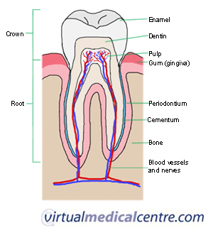
Each tooth consists of a crown and one or more roots.
The crown is the functional part that is visible above the gum. The root is the unseen portion that supports and fastens the tooth in the jawbone. The clear outer layer of the crown is the Enamel, the hardest substance in the human body. The outer layer of the roor is cementum, a bonelike substance that anchors the tooth to the jawbone. Directly beneath the outer layer is dentin, a hard, mineral material that is similar to human bone, only stronger. Dentin surrounds and protects the pulp, or core of the tooth. Pulp contains blood vessels, which carry oxygen and nutrients to the tooth, and nerves, which transmit pain and temperature sensations to the brain.
The muscular tongue helps mix the food with saliva and the enzymes it contains.
When you swallow, the lump of chewed food, now called a bolus, passes into the pharynx.
The pharynx serves both the respiratory system and the digestive system. It connects the mouth to the rest of the digestive tract and, also connects the mouth and nose to the rest of the respiratory system. To prevent food or liquid from entering the trachea (windpipe), the epiglottis (a small flap of tissue) closes over the opening of the larynx (voice box) during swalowing (deglutition).

miércoles, 8 de diciembre de 2010

The digestive system

Nutrients in the foods you eat are needed by the cells of your body, we eat foods to obtein nutrients. But:
What organs and processes break down the foofs into nutrients that the body can use for fuel?
What organs and processes let the nutrients enter your bloodstream so they can travel to the cells where they are needed?
The basic processes involved are digestion and absorption. The organs involved are the organs of the digestive system.
Organs that make up the digestive system are shown in figure. Most of the organs form the gastrointestinal tract. Other digestive organs are called accessory organs.


Organs of the digestive system.
1. Gastrointestinal Tract
The gastrointestinal (GI) tract is a long tube that connects the mouth with the anus. It is more than 9 meters long in adults. The upper GI tract includes the mouth, pharynx, esophagus, and stomach. The lower GI tract includes the small and large intestines. Food enters the mouth, passes through the upper and lower GI tracts, and then exits the body as feces through the anus.

The organs of the GI tract are covered by two layers of muscles that enable peristalsis. Peristalsis is a rapid, involuntary, wave-like contraction of muscles. It pushes food through the GI tract.
The inside of GI tract is lined with mucous membranes. Mucous membranes can secrete and absorb substances. The ability to secrete and absorb substances is necessary for the functions of the digestive system.
2. Accessory Organs of the Digestive System
They are additional organs that play important roles in digestion. Food does not pass through them, but they make substances needed for digestion. The accessory organs are the salivary glands, the liver and the pancreas.

  • The salivary glands are found in and around your mouth and throat. We call the major salivary glands the parotid, submandibular, and sublingual glands.They all secrete saliva into your mouth .
  • The liver is a large organ next to the stomach. It produces digestive substances that are carried by tubes,to the small intestine and gall bladder. The gall bladder is a small, pear-shaped structure below the liver. It stores substances (bile) from the liver until they are needed by the small intestine.
  • The pancreas is a gland below the stomach. It produces digestive substances that are carried by a duct to the small intestine.