Mostrando entradas con la etiqueta skeleton. Mostrar todas las entradas

martes, 17 de mayo de 2011

Skeleton vocabulary

I'm sure some of you were missing the post about the vocabulary:


  • Bone marrow Soft connective tissue found inside many bones; site of blood cell formation.

  • Cartilage Smooth covering found at the end of bones; made of tough collagen protein fibers; creates smooth surfaces for the easy movement of bones against each other.

  • Compact bone The dense, hard outer layer of a bone.

  • Joint Point at which two or more bones meet.

  • Ligaments Fibrous tissue that connects bones to other bones; made of tough collagen fibers.

  • Movable joint Most mobile type of joint; the most common type of joint in the body.

  • Skeletal system Body system that is made up of bones, cartilage, and ligaments.

  • Skeleton Sturdy scaffolding of bones and cartilage that is found inside vertebrates.

  • Spongy bone Lighter and less dense than compact bone; found toward the center of the bone.

  • Sprain A ligament injury; usually caused by the sudden overstretching of a joint which causes tearing.

martes, 10 de mayo de 2011

Joints and How they move

A joint is a point at which two or more bones meet. There are three types of joints in the body:
Fixed joints is a joint between two bones that doesnt move . Many of the joints in your skull are fixed joints. The skull plates don't move together or against each other, but they are connected or fused
Partly movable joints allow only a little movement..Your backbone has partly movable joints between the vertebrae. They are connected to each other by pads of cartilageand ligaments. They can only move a small amount
Movable joints allow movement and provide mechanical support for the body. Movable joints are the most common type of joint in your body. Your fingers, toes, hips, elbows, and knees all have movable joints. The surfaces of bones at movable joints are covered with a smooth layer of cartilage. The space between the bones in a movable joint is filled with a liquid called synovial fluid. The fluid lubricates and cushions the bones when they move at the joint. Ligaments help provide their stability and muscles contract to produce movement.
The knee joint is the largest joint in the body, consisting of 4 bones- the femur (thigh bone), the tibia (shin bone), fibula (outer shin bone) and patella (kneecap)- and an extensive network of ligaments and muscles..
There is a joint capsule that is a thick ligamentous structure that surrounds the entire knee. Inside this capsule is a specialized membrane known as the synovial membrane which provides nourishment to all the surrounding structures. The capsule itself is strengthened by the surrounding ligaments.
The stability of the knee owes greatly to the presence of its ligaments. Each has a particular function in helping to maintain optimal knee stability in a variety of different positions. There are two Collateral Ligaments ( inner and outer side) and two Cruciate ligaments.
Each knee joint has two crescent-shaped cartilage menisci. These lie on the medial (inner) and lateral (outer) edges of the upper surface of the tibia bone. They are essential components, acting as shock absorbers for the knee as well as allowing for correct weight distribution between the tibia and the femur.

domingo, 8 de mayo de 2011

Appendicular Skeleton


Appendicular Skeleton The appendicular skeleton consists of the bones of>the upper and lower extremities.
UPPER EXTREMITY
The upper extremity consists of the bones of the shoulder (pectorla girdle), the arm, the forearm, the wrist, and the hand.

  • Clavicle.The clavicle (commonly called the collar bone) The clavicle and the scapula forms the pectoral girdle
  • Scapula.The scapula is a triangular bone that lies in the upper part of the back
  • Humerus.—The humerus is the longest bone of the upper extremity and is often called the arm bone It articulates with the pectoral girdle to form the shoulder joint, and with the bones of the forearm to form the elbow
  • Radius and Ulna.—When the arm is in the anatomical position with the palm turned forward, the radius is on the lateral (thumb) side and the ulna is on the medial (little finger) side of the forearm
  • Carpal.There are eight carpal bones, arranged in two rows, forming the wrist. Metacarpal.The metacarpal bones are in the palm
  • Phalanges.The small bones of the fingers are called phalanges, and each one of these bones is called a phalanx. Each finger has three phalanges, except the thumb (which has two)
LOWER EXTREMITY.—The lower extremity includes the bones of the hip (pelvic girdle), thigh, leg, ankle, and foot
  • Pelvic bones.— Commonly known as the hip, is a large, irregularly shaped bone composed of three parts: the ilium, ischium, and pubis
  • Femur.The femur, or thigh bone, is the longest bone in the body (fig. 1-24).
  • Patella.The patella is a small oval-shaped bone overlying the knee joint. It is enclosed within the tendon of the quadriceps muscle of the thigh.
  • Tibia.The tibia, or shin bone, is the larger of the two leg bones and lies at the medial side.
  • Fibula.The fibula, the smaller of the two leg bones, is located on the lateral side of the leg, parallel to the tibia.
  • Tarsus.—The tarsus, or ankle, is formed by seven tarsal bones
  • Metatarsus.—The sole and instep of the foot is called the metatarsus and is made up of five metatarsal bones
  • The phalanges are the bones of the toes and are similar in number, structure, and arrangement to the bones of the fingers

Axial Skeleton

DIVISIONS OF SKELETON
The human skeleton can be divided into two main parts; the first part is the axial skeleton and second part is the appendicular skeleton.
Axial Skeleton:
The axial skeleton consists of the skull, thevertebral column, and the thorax.

SKULL.— The skull is the bony structure in the head. It supports the structures of the face and forms a cavity for the brain.The skull is composed of two parts: the cranium and the mandible

Cranial Bones.—The cranium is formed by eight major bones, most of which are in pairs.
The frontal bone forms the forehead and the nasal cavity.
The parietal bones form the roof of the skull.The temporal bones help form the sides and base of the skull and also house the auditory and hearing organs.
The occipital bone forms part of the base and back of the skull, and contains a large hole called the foramen magnum
The sphenoid bone is situated on the base of the skull
The ethmoid bone is located in front of the sphenoid
Facial Bones.— : 13 immovable bones and a movable lower jawbone
The maxillary bones form the upper jaw, the anterior roof of the mouth, the floors of the orbits, and the sides and floor of the nasal cavity
The lower jawbone is called the mandible

VERTEBRAL (SPINAL) COLUMN
The vertebral column consists of 24 movable or true vertebrae; the sacrum; and the coccyx or tail bone. The bodies of the vertebrae are separated from each other by intervertebral discs

The spinal column is divided into five regions in the following order:
Cervical (neck), seven vertebrae
Thoracic (chest) twelve vertebrae that articulate with the ribs
Lumbar five lumbar vertebrae located in the small of the back, these vertebrae are the larger and stronger segments of the vertebral column
Sacrum is the triangular bone immediately below the lumbar vertebrae. It is composed of five fused vertebrae The sacrum is connected on each side with the hip bone and with the coccyx to form the posterior wall of the pelvis.
Coccyx is the tailbone. It have four fused vertebrae
THORAX.
The skeleton of the thorax includes 12 ribs on each side articulates posteriorly with the thoracic vertebrae and the sternum in the front upper half of the chest wall.
ribs 1 to 7 are connected to the sternum' by their costal cartilages and are called true ribs
ribs 8 to 10 are termed false ribs. They join the costal cartilage immediately above.by means of their costal cartilages.
ribs 11 and 12, which are free, are known as floating ribs.

Structure of Bones

Bones are rigid organs that are made up of two main types of bone tissue:
Compact bone makes up the dense outer layer of bones.
Spongy bone is lighter and less dense than compact bone, and is found toward the center of the bone. The tough, shiny, white membrane that covers all surfaces of bones is called the periosteum
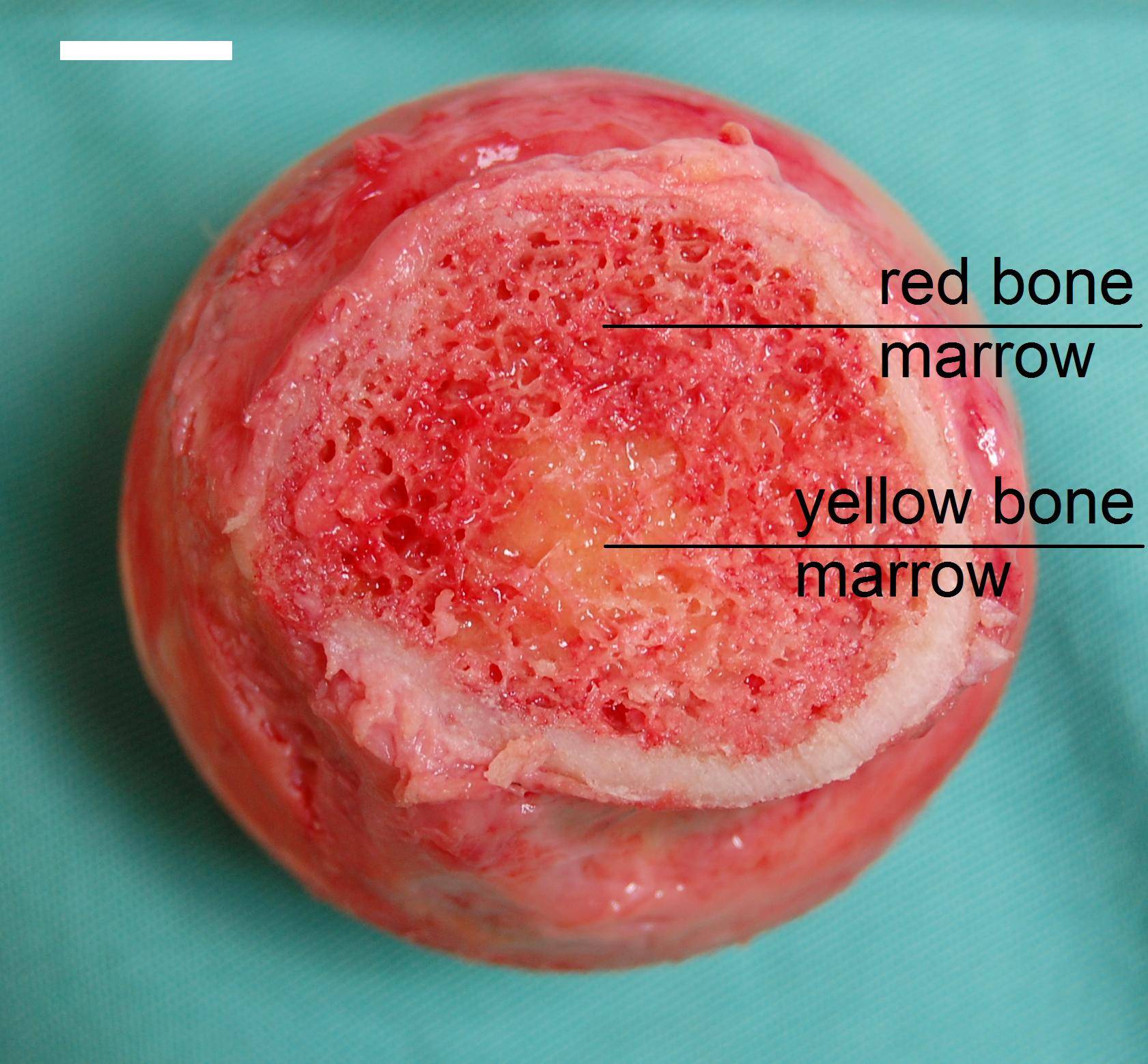
Many bones also contain in their innermost part a soft connective tissue called bone marrow. There are two types of bone marrow: red marrow and yellow marrow. Red marrow makes blood cells. Yellow marrow consists of fat cells although it will have a different role to that of normal fat cells
The bones of newborn babies contain only red marrow. As children get older, thei red marrow is replaced by yellow marrow. In adults, red marrow is found mostly in the bones of the skull, the ribs, and pelvic bones.

Classification of bones
There are four main types of bones in the human body. They can be long, short, flat, or irregular. Identifying a bone as long, short, flat, or irregular is based on the shape of the bone not the size of the bone.
Long bone are longer than wide . They consist of a long shaft (diaphysis) with two bulky ends or extremities(epiphyses). The structure of a long bone is shown in Figure. They are primarily compact bone but may have a large amount of spongy bone at the ends or extremities. Long bones include femur, tibia, and fibula of the leg, the humerus, radius, and ulna of the arm and the phalanges of the fingers and toes.
Short bones are roughly cube shaped with vertical and horizontal dimensions approximately equal. They consist primarily of spongy bone, which is covered by a thin layer of compact bone. Short bones include the bones of the wrist and ankle.
Flat bones are thin, flattened, and usually curved. Most of the bones of the cranium are flat bones.
Irregular bones are not in any of the above three categories. They are primarily spongy bone that is covered with a thin layer of compact bone. The vertebrae and some of the bones in the skull are irregular bones.
In this video you can watch the different types of bones:

Skeleton

Humans are vertebrates, which are animals that have a backbone. The sturdy scaffolding of bones and cartilage that is found inside vertebrates is called a skeleton. The adult human skeleton has about 206 bones, some of which are named in Figure.
The skeletal system is made up of bones, cartilage, and ligaments.
Bones are not only the hard mineral part as the remains that you might see in a museum. Living bones are full of life. They contain nerves, blood vessels and many different types of tissues
Cartilage is found at the end of bones and is made of tough protein fibers called collagen. Cartilage creates smooth surfaces for the movement of bones that are next to each other, like the bones of the knee.
Ligaments are made of tough protein fibers and connect bones to each other.
Functions of Bones
Your skeletal system gives shape and form to your body, but it is also important in other homeostatic functions. The main functions of the skeletal system are:
  • Support The skeleton supports the body against the pull of gravity. The large bones of the lower limbs support the trunk when standing.
  • Protection The skeleton provides a framework that supports and protects the soft organs of the body. For example, the skull surrounds the brain to protect it from injury. The bones of the rib cage help protect the heart and lungs.
  • Movement Bones work together with muscles as simple mechanical lever systems to move the body.
  • Making Blood Cells Blood cells are made mostly inside certain types of bones.
  • Storage Bones store calcium. They contain more calcium than any other organ does. Calcium is released by the bones when blood levels of calcium drop too low. The mineral phosphorus is also stored in bones.
As in other units in this link Kidshealth you can read and listen information about this subject, and finally a video with a reviw of all about skeleton.