Belen wants to help us to learn the circulatory system, so she sent me this video from youtube. Maybe, you will improve your understanding of it with a little bit of music.
As this post is a little different from the others, here you have two interesting posts, at least for me, related with music. I discovered them in the blog of music of Ciudad Real CPR
Eric Whitacre's Virtual Choir - 'Lux Aurumque'
World Science Festival 2009: Bobby McFerrin Demonstrates the Power of the Pentatonic Scale
lunes, 31 de enero de 2011

jueves, 27 de enero de 2011
The Composition of Blood

Blood is a fluid connective tissue. It circulates around the body through the blood vessels by the pumping action of the heart.
Blood accounts for about 7% of the human body weight. The average adult has a blood volume of roughly 5 liters, composed of:
Blood accounts for about 7% of the human body weight. The average adult has a blood volume of roughly 5 liters, composed of:
- A fluid called plasma where are the blood cells and other substances.
- Several kinds of blood cells. Within the blood plasma, are:
- Erythrocytes (red blood cells)
- Leukocytes (white blood cells)
- Thrombocytes (platelets)
The cells that make up the blood can be seen in Figure
 Plasma
Plasma is the golden-yellow liquid part of the blood. Plasma is 90% water and 10% dissolved materials including proteins, glucose, ions, hormones, and gases.
Red Blood Cells
Red blood cells, also known as erythrocytes, are flattened, doubly concave cells that carry oxygen. There are about 4 to 6 million cells per cubic millimeter of blood. Red blood cells are continuously made in the red bone marrow of long bones, ribs, skull, and vertebrae. Each red blood cell lives for only 120 days, after which they are destroyed in liver and spleen.
Mature red blood cells do not have a nucleus or other organelles. They contain the protein hemoglobin which iron and this protein gives blood its red color
White Blood Cells
White blood cells, also known as leukocytes, are generally larger than red blood cells,. They have a nucleus. White blood cells make up less than one percent of the blood's volume. They are made from stem cells in bone marrow. They take part in the immune response. There are five types of white blood cells but the most important are:
Monocytes enter the tissue fluid by squeezing through capillary walls and transform into Macrophages that phagocytose (swallow) cell debris and. bacteria or viruses
Neutrophils also swallow foreign bodies.
Lymphocytes fight infection. They produce antibodies, proteins that travel in blood to identify and neutralize foreign objects, such as bacteria and viruses
Macrophage showing cytoplasmic extensions that allow it to swallow particles or pathogens. In the image here, a mouse macrophage stretches its arms to engulf two particles at once.
Platelets
Platelets, also known as thrombocytes, are small, regularly-shaped clear cell fragments. They are important in blood clotting. Platelets carry chemicals essential to blood clotting. This clot stops more blood from leaving the body when damage to wall of blood vessels occurs
 Cells of the blood. From left to right: Red blood cell, platelet, white blood cell.
The Components of Blood
Pulmonary and Systemic Circulations
The double circulatory system of blood flow refers to the separate systems of pulmonary circulation and the systemic circulation in amphibians, birds and mammals, including humans. Pulmonary Circulation
The pulmonary circulation is the portion of the cardiovascular system which carries oxygen-poor (deoxygenated) blood away from the heart, to the lungs, and returns oxygenated blood back to the heart. As shown in Figure, deoxygenated blood from the body leaves the right ventricle through the pulmonary arteries, which carry the blood to each lung. The pulmonary arteries are the only arteries that carry deoxygenated blood. In the lungs, red blood cells release carbon dioxide and pick up oxygen during respiration. The oxygenated blood then leaves the lungs through the pulmonary veins, which return it to the left side of the heart, and complete the pulmonary cycle. In the following scheme of the general body circulation, pulmonary circulation is in the right side and the systemic circulation is on the left side.

Systemic Circulation
The systemic circulation is the portion of the cardiovascular system which carries oxygenated blood away from the heart, to the body, and returns deoxygenated blood back to the heart. Oxygenated blood from the lungs leaves the left ventricle through the aorta, from where it is distributed to the body's organs and tissues, which absorb the oxygen, through a complex network of arteries and capillaries. The deoxygenated blood is then collected by veins and then into the inferior and superior venae cavae, which return it to the right heart, completing the systemic cycle. The blood is then re-oxygenated through the pulmonary circulation before returning again to the systemic circulation.
Just like every other organ in the body, the heart needs its own blood supply, which it gets through the coronary arteries that branch directly from the aorta, just above the heart. They deliver oxygen-rich blood to the heart
Pulmonary CirculationThe pulmonary circulation is the portion of the cardiovascular system which carries oxygen-poor (deoxygenated) blood away from the heart, to the lungs, and returns oxygenated blood back to the heart. As shown in Figure, deoxygenated blood from the body leaves the right ventricle through the pulmonary arteries, which carry the blood to each lung. The pulmonary arteries are the only arteries that carry deoxygenated blood. In the lungs, red blood cells release carbon dioxide and pick up oxygen during respiration. The oxygenated blood then leaves the lungs through the pulmonary veins, which return it to the left side of the heart, and complete the pulmonary cycle. In the following scheme of the general body circulation, pulmonary circulation is in the right side and the systemic circulation is on the left side.
Systemic Circulation
The systemic circulation is the portion of the cardiovascular system which carries oxygenated blood away from the heart, to the body, and returns deoxygenated blood back to the heart. Oxygenated blood from the lungs leaves the left ventricle through the aorta, from where it is distributed to the body's organs and tissues, which absorb the oxygen, through a complex network of arteries and capillaries. The deoxygenated blood is then collected by veins and then into the inferior and superior venae cavae, which return it to the right heart, completing the systemic cycle. The blood is then re-oxygenated through the pulmonary circulation before returning again to the systemic circulation.
| Pulmonary Circulation | Systemic Circulation |
It is a shorter circulation. | It is a larger circulation. The circulation is between heart and remaining parts of the body except lungs. Blood is pumped by left part of the heart and received by the right part. It pumps oxygenated blood to different parts of the body. It brings back deoxygenated blood to the heart. |
Just like every other organ in the body, the heart needs its own blood supply, which it gets through the coronary arteries that branch directly from the aorta, just above the heart. They deliver oxygen-rich blood to the heart
Blood Vessels
Arteries are the large, muscular vessels that carry blood away from the heart toward the organs.. Arteries have thicker walls than veins. The elastic qualities of artery walls allow them to carry pressurized blood from the heart while maintaining blood pressure.
Arteries branch into smaller arteries and finally the capillaries that carry nutrients to the body’s cells and tissues.

Veins are vessels that carry blood from the organs toward the heart. The walls of veins have one-way valves that prevent blood from flowing backward and pooling in the legs, feet, arms or hands due to the pull of gravity. The location of veins can vary from person to person.
Capillaries are the smallest of the body's blood vessels, measuring 5-10 μm in diameter. They connect smoller arteries (arterioles) and smoller veins (venules), and they are important for the exchange of oxygen, carbon dioxide, and other substances between blood and body cells. The walls of capillaries are made of only a single layer of endothelial cells. This layer is so thin that molecules such as oxygen, water and lipids can pass through them by diffusion and enter the body tissues.
In general the term arterial blood is used to describe blood high in oxygen and venous blood to describe blood low in oxygen, although the pulmonary arteries carry deoxygenated blood and blood flowing in the pulmonary vein is rich in oxygen. (...)
Blood Vessels and Blood Pressure
Blood pressure refers to the force exerted by circulating blood on the walls of blood vessels. The pressure of the circulating blood gradually decreases as blood moves from the arteries, capillaries, and veins. The term "blood pressure" generally refers to arterial pressure, which is the pressure in the larger arteries that take blood away from the heart. Arterial pressure results from the force that is applied to blood by the contracting heart, where the blood “presses” against the walls of the arteries. During each heartbeat, BP varies between a maximum (systolic) and a minimum (diastolic) pressure.
The systolic arterial pressure is defined as the peak pressure in the arteries, which occurs systole( contraction); the diastolic arterial pressure is the lowest pressure when the heart is in diastole (relaxation).
the healthy ranges for arterial pressure are:
• Systolic: less than 120 mm Hg
• Diastolic: less than 80 mm Hg
Factors such as age, gender and race influence blood pressure values. Pressure also varies with exercise, emotional reactions, sleep, stress, nutritional factors, drugs, or disease. Hypertension is a condition in which a person’s blood pressure is chronically high.
This link to Discovery Health you have a video with a good explanation of blood pressure and this video is also interesting
martes, 25 de enero de 2011
The Heartbeat
- As we saw in the last post, the heart is a four-chambered organ consisting of right and left halves. Two of the chambers, the left and right atria, are entry-points into the heart, while the other two chambers, the left and right ventricles, are responsible for contractions that send the blood through the circulation.
The circulation is split into the pulmonary and systemic circulation. The right ventricle's role is to pump deoxygenated blood into the pulmonary circulation through the pulmonary artery. The left ventricle's role is to pump now oxygenated blood into the systemic circulation through the aorta.
The average human heart, beating at 72 beats per minute, will beat approximately 2.5 billion times during an average 66 year lifespan. Sometime the heart can beat fast, this is called Tachycardia. It happen when you make exercise or are in danger.
The heartbeat is made up of two parts;.
Systole is the contraction of the heart chambers, which drives blood out of the chambers.
Diastole is the period of time when the heart relaxes after contraction.
In the figura you have the cardiac cycle :

- Atrial diastole. The atria were in diastole and blood from the superior and inferior vena cava (rigth side) and pulmonary veins (left side) flows into the atria slowly to fill them and begin the cycle.
- Atrial systole. This phase involves the contraction of the 2 atria, pushing the blood into the respective ventricles. There is no back flow of blood due to the presence of the atrioventricular (AV) valves ( bicuspid valve – left and tricuspid valve -right) . The bicuspid valve is supported by tendons which look rather like the strings of a parachute.
- Ventricular systole. The thick muscular walls of the ventricles contract.. This begins alongside the end of auricular diastole. The pressure on the blood in the ventricles increases. The atrioventricular valves close rapidly to prevent the backward flow of blood into the auricles.
As the pressure in the ventricle increases, the semilunar valves are opened and blood enters the arteries. From the right ventricle, the deoxygenated blood enters the pulmonary artery. From the left ventricle, the oxygenated blood enters the aorta, to be taken to all body parts. - Ventricular diastole.Ventricular systole is followed by ventricular diastole. The atria are already in diastole, so all the chambers of the heart are in diastole. As the pressure in the ventricles decreases to prevent the backward flow of blood, the semilunar valves close rapidly.
The sound of the heart valves shutting causes the heart sounds, or a heartbeat. The closing of the mitral and tricuspid valves (known together as the atrioventricular valves) at the beginning of ventricular systole cause the first part of the "lub-dub" sound made by the heart as it beats. The second part of the "lub-dub" is caused by the closure of the aortic and pulmonic valves at the end of ventricular systole. As the left ventricle empties, its pressure falls below the pressure in the aorta, and the aortic valve closes. Similarly, as the pressure in the right ventricle falls below the pressure in the pulmonary artery, the pulmonic valve closes.
Here you have some videos to get a better knowledge of the heartbeat.
The heart
The heart is usually found in the left to middle of the chest with the largest part of the heart slightly to the left. It is about the size of a fist. The heart is surrounded by the lungs.
It is divided into four chambers, the two upper atria and the two lower ventricles. Atria (singular, atrium) are the thin-walled blood collection chambers of the heart. Atria pump the blood into the ventricles. Ventricles are the heart chambers which collect blood from the atria and pump it out of the heart. The four chambers of the heart are shown in Figure. Each of the four chambers of the heart have a specific job, these are:
• The right atrium receives oxygen-poor (deoxygenated) blood from the body this blood enters from the superior vena cava and the inferior vena cava
• The right ventricle pumps oxygen-poor blood through the pulmonary arteries and toward the lungs. In the lungs, carbon dioxide is released from the blood and oxygen is picked up.
• The left atrium receives oxygen-rich (oxygenated) blood from the lungs through the pulmonary veins.
• The left ventricle pumps oxygen-rich blood out of the heart to the rest of the body through the aorta.
On both sides, the lower ventricles are thicker and stronger than the upper atria. The muscle wall surrounding the left ventricle is thicker and stronger than the wall surrounding the right ventricle because the left ventricle needs to exert enough force to pump the blood through the body. The right ventricle only needs to pump the blood as far as the lungs, which does not require as much contractile force.
Valves in the heart maintain the flow of blood by opening and closing in one direction only. Blood can move only forward through the heart, and is prevented from flowing backward by the valves. Such movement of the blood is called unidirectional flow. There are four valves of the heart:
• The two atrioventricular (AV) valves ensure blood flows from the atria to the ventricles, and not the other way. The AV valve on the right side of the heart is called the tricuspid valve, and the one on the left of the heart is called the mitral, or bicuspid valve.
• The two semilunar (SL) valves are present in the arteries leaving the heart, and they prevent blood flowing back from the arteries into the ventricles.
It is divided into four chambers, the two upper atria and the two lower ventricles. Atria (singular, atrium) are the thin-walled blood collection chambers of the heart. Atria pump the blood into the ventricles. Ventricles are the heart chambers which collect blood from the atria and pump it out of the heart. The four chambers of the heart are shown in Figure.
Each of the four chambers of the heart have a specific job, these are:• The right atrium receives oxygen-poor (deoxygenated) blood from the body this blood enters from the superior vena cava and the inferior vena cava
• The right ventricle pumps oxygen-poor blood through the pulmonary arteries and toward the lungs. In the lungs, carbon dioxide is released from the blood and oxygen is picked up.
• The left atrium receives oxygen-rich (oxygenated) blood from the lungs through the pulmonary veins.
• The left ventricle pumps oxygen-rich blood out of the heart to the rest of the body through the aorta.
On both sides, the lower ventricles are thicker and stronger than the upper atria. The muscle wall surrounding the left ventricle is thicker and stronger than the wall surrounding the right ventricle because the left ventricle needs to exert enough force to pump the blood through the body. The right ventricle only needs to pump the blood as far as the lungs, which does not require as much contractile force.
Valves in the heart maintain the flow of blood by opening and closing in one direction only. Blood can move only forward through the heart, and is prevented from flowing backward by the valves. Such movement of the blood is called unidirectional flow. There are four valves of the heart:
• The two atrioventricular (AV) valves ensure blood flows from the atria to the ventricles, and not the other way. The AV valve on the right side of the heart is called the tricuspid valve, and the one on the left of the heart is called the mitral, or bicuspid valve.
• The two semilunar (SL) valves are present in the arteries leaving the heart, and they prevent blood flowing back from the arteries into the ventricles.
domingo, 23 de enero de 2011
The circulatory system
After seeing digestive and respiratory system, the next one is the cardiovascular system. It has a pretty important function in the nutrition role. As you already know, every cell in your body depends on your cardiovascular system. It keeps all of your cells supplied with nutrients from the intestine (digestive system) and oxygen from the lungs (respiratory system). It also removes their waste products, carbon dioxide to the lungs and the nitrogenous wastes to the kidneys.
Actually the circulatory system has many jobs, but we can cosider three main functions:
• Transport of nutrients, oxygen, and hormones to cells throughout the body and removal of metabolic wastes (carbon dioxide, nitrogenous wastes, and heat).
• Protection of the body by white blood cells and antibodies that circulate in the blood and defend the body against foreign microbes and toxins. Clotting mechanisms are also present that protect the body from blood loss after injuries.
• Regulation of body temperature and fluid pH...
This video can be a good help to begin with the circulatory system.
The cardiovascular system shown in Figure is made up of: The heart
It pushes the blood around your body through the blood vessels. The heart is made of cardiac muscle. Blood is collected in the heart and pumped out to the lungs, where it releases carbon dioxide and picks up oxygen before it is pumped to the rest of the body.
The blood vessels
Their job is to channel the blood around the body. There are three main types of blood vessels in the body; arteries, veins, and capillaries
Arteries are blood vessels that carry blood away from the heart. Further from the heart, arteries form smaller arteries. These smaller arteries branch into smaller vessels. The smaller blood vessels help to bring nutrients and oxygen and take away waste from body tissues.
Capillaries are the tiniest blood vessels in the body located within the tissues of the body. They transport blood from the arteries to the veins. The walls of capillaries are only a single layer of cells thick. Oxygen, carbon dioxide, nutrients, and wastes are exchanged through their thin walls. Capillaries are so narrow that blood cells must move in single file through them.
Veins are blood vessels that carry back the blood from the different regions of the body to the heart.
The blood
Blood is a body fluid that is a type of connective tissue. Blood is made of blood cells, and a fluid called plasma. The main types of cells found in blood are red blood cells and white blood cells.
Finally remember you something you studied last course. The cardiovascular system of humans is closed. That means the blood never leaves the large loop of blood vessels in which it travels. Other animals such as invertebrates have open circulatory systems, in which their blood can leave the blood vessels.
Actually the circulatory system has many jobs, but we can cosider three main functions:
• Transport of nutrients, oxygen, and hormones to cells throughout the body and removal of metabolic wastes (carbon dioxide, nitrogenous wastes, and heat).
• Protection of the body by white blood cells and antibodies that circulate in the blood and defend the body against foreign microbes and toxins. Clotting mechanisms are also present that protect the body from blood loss after injuries.
• Regulation of body temperature and fluid pH...
This video can be a good help to begin with the circulatory system.
The cardiovascular system shown in Figure is made up of:
The heartIt pushes the blood around your body through the blood vessels. The heart is made of cardiac muscle. Blood is collected in the heart and pumped out to the lungs, where it releases carbon dioxide and picks up oxygen before it is pumped to the rest of the body.
The blood vessels
Their job is to channel the blood around the body. There are three main types of blood vessels in the body; arteries, veins, and capillaries
Arteries are blood vessels that carry blood away from the heart. Further from the heart, arteries form smaller arteries. These smaller arteries branch into smaller vessels. The smaller blood vessels help to bring nutrients and oxygen and take away waste from body tissues.
Capillaries are the tiniest blood vessels in the body located within the tissues of the body. They transport blood from the arteries to the veins. The walls of capillaries are only a single layer of cells thick. Oxygen, carbon dioxide, nutrients, and wastes are exchanged through their thin walls. Capillaries are so narrow that blood cells must move in single file through them.
Veins are blood vessels that carry back the blood from the different regions of the body to the heart.
The blood
Blood is a body fluid that is a type of connective tissue. Blood is made of blood cells, and a fluid called plasma. The main types of cells found in blood are red blood cells and white blood cells.
Finally remember you something you studied last course. The cardiovascular system of humans is closed. That means the blood never leaves the large loop of blood vessels in which it travels. Other animals such as invertebrates have open circulatory systems, in which their blood can leave the blood vessels.
martes, 18 de enero de 2011
Lung volume
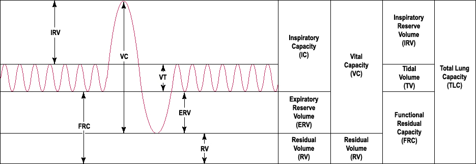
Lung volumes and lung capacities refer to the volume of air associated with different phases of the respiratory cycle. Lung volumes are directly measured. Lung capacities are inferred from lung volumes.
The average total lung capacity of an adult human male is about 6 litres of air, but only a small amount of this capacity is used during normal breathing.
The breathing mechanism is called respiration. Tidal breathing is normal, resting breathing; the tidal volume is the volume of air that is inhaled or exhaled in a single such breath. It's 0.5 litres
An average human breathes some 12-20 times per minute.
Several factors affect lung volumes; some can be controlled and some cannot. Lung volumes can be measured using the following terms:
A person who is born and lives at sea level will develop a slightly smaller lung capacity than a person who spends their life at a high altitude. This is because the partial pressure of oxygen is lower at higher altitude which, as a result means that oxygen less readily diffuses into the bloodstream. In response to higher altitude, the body's diffusing capacity increases in order to process more air.
When someone living at or near sea level travels to locations at high altitudes (eg. the Andes, Tibet, the Himalayas, etc.) that person can develop a condition called altitude sickness because their lungs remove adequate amounts of carbon dioxide but they do not take in enough oxygen. (In normal individuals, carbon dioxide is the primary determinant of respiratory drive.)
The average total lung capacity of an adult human male is about 6 litres of air, but only a small amount of this capacity is used during normal breathing.
The breathing mechanism is called respiration. Tidal breathing is normal, resting breathing; the tidal volume is the volume of air that is inhaled or exhaled in a single such breath. It's 0.5 litres
Average lung volumes in healthy adults | ||
Volume | Men (Value-litres) | Women(Value-litres) |
Inspiratory reserve volume | 3.3 | 1.9 |
Tidal volume | 0.5 | 0.5 |
Expiratory reserve volume | 1.0 | 0.7 |
Residual volume | 1.2 | 1.1 |
| Volume | Men | Women | Derivation /td> |
| Vital capacity | 4.8 | 3.1 | IRV+TV+ERV |
| Inspiratory capacity | 3.8 | 2.4 | IRV+TV |
| Functional residual capacity | 2.2 | 1.8 | ERV+RV |
| Total lung capacity | 6.0 | 4.2 | IRV+TV+ERV+RV |
An average human breathes some 12-20 times per minute.
Several factors affect lung volumes; some can be controlled and some cannot. Lung volumes can be measured using the following terms:
| Larger volumes | Smaller volumes |
| taller people | shorter people |
| non-smokers | smokers |
people who live at higher altitudes | people who live at lower altitudes |
When someone living at or near sea level travels to locations at high altitudes (eg. the Andes, Tibet, the Himalayas, etc.) that person can develop a condition called altitude sickness because their lungs remove adequate amounts of carbon dioxide but they do not take in enough oxygen. (In normal individuals, carbon dioxide is the primary determinant of respiratory drive.)
domingo, 16 de enero de 2011
Respiratory system vocabulary
alveoli:Little "sacs" at the end of the bronchioles where most of the gas exchange occurs.
asthma:A chronic illness in which the bronchioles are inflamed and become narrow.
bronchitis:An inflammation of the bronchi.
diaphragm:A sheet of muscle that extends across the bottom of the rib cage. When the diaphragm contracts the chest volume gets larger and the lungs take in air; when the diaphragm relaxes, the chest volume gets smaller and air is pushed out of the lungs.
emphysema: A chronic lung disease caused by loss of elasticity of the lung tissue.
epiglottis:A flap of connective tissue that closes over the trachea when food is swallowed to prevent choking or inhaling food.
exhalation:Pushing air out of the body through the nose or mouth.
external respiration:The movement of oxygen into the body and carbon dioxide out of the body.
inhalation:Taking air into the body through the nose and mouth.
internal respiration:The exchange of gases between the blood and the cells of the body.
larynx:Found just below the point at which the pharynx splits into the trachea and the esophagus. Your voice comes from your larynx; air from the lungs passes across thin membranes in the larynx and produces sound; also called the voicebox.
lung cancer:A disease where the cells that line the lungs grow out of control; the growing mass of cells pushes into nearby tissues and can affect how these tissues work.
pathogen: An organism that causes disease in another organism; certain bacteria, viruses, and fungi are pathogens of the respiratory system.
pharynx:A long tube that is shared with the digestive system; both food and air pass through the pharynx.
pneumonia:An illness in which the alveoli become inflamed and flooded with fluid.
respiratory disease
trachea:A long tube that leads down to the chest where it divides into the right and left bronchi in the lungs; also called the windpipe.
tuberculosis (TB): A common and often deadly infectious disease caused by a type of bacterium called mycobacterium.
The Journey of a Breath of Air
When you breath in, oxygen is drawn in through the nose or mouth and down into the lungs. The oxygen then passes across the thin lining of the capillaries and into the blood. The oxygen molecules are carried to the body cells by the blood. Carbon dioxide from the body cells is carried by the blood to the lungs where it is released into the air.
Breathing is only part of the process of delivering oxygen to where it is needed in the body. Gas exchange occurs in the alveoli by passive diffusion of gases between the alveoli and the blood in the capillaries of the lungs.

Diffusion is the movement of substances from an area of higher concentration to an area of lower concentration. The concentration of O2 in the alveoli is at a higher level than in the blood and the concentration of CO2 in the alveoli is at a lower lever than in the blood. O2 molecules diffuse across the thin walls of the alveoli and capillaries and into the blood. Carbon dioxide (CO2) moves out of the blood and into the alveoli in a similar way
The pulmonary artery carries the blood traveling to the lungs and alveoli. Upon reaching the alveoli the blood picks up the inhaled oxygen and at the same time releases the carbon dioxide that needs to be expelled. The pulmonary veins then carry the oxygenated blood to the heart to be pumped through the aorta and around the body. The oxygenated blood travels to the smaller arteries and finally to the capillaries where gas exchange occurs.
The oxygen molecules move out of the capillaries and into the body cells. It is used by the cells to Cellular respiration, the process of breaking down glucose to release energy .
C6H12O6 + 6O2 → 6H2O + 6 CO2 + ENERGY
The waste products of cellular respiration include carbon dioxide and water. The carbon dioxide molecules move out of the cells and into the capillaries that surround the cells. The carbon dioxide is removed from the body by the lungs.
Can you believe it?
Three post on the respiratory system and no video yet, can you believe it?. In this post just in case you are missing the videos, here you have some of them.
The first one is for kids but very clear and easy to understand.
The second one is a reviw of all the parts and organs of the respiratory system.
In the last one of this post you can understand the breathing movement.
Good work and until the next post.
The first one is for kids but very clear and easy to understand.
The second one is a reviw of all the parts and organs of the respiratory system.
In the last one of this post you can understand the breathing movement.
Good work and until the next post.
sábado, 15 de enero de 2011
How we breathe
Most of the time, you breathe without thinking of it. Breathing is mostly an involuntary action that is controlled by a part of your brain that also controls your heart beat. If you swim or sing, by instance, you know you can also control your breathing.
The taking in and expelling out of air is done by two movements:Inspiration and expiration. Inspiration: breathing in (a.k.a., inhalation)
Taking air into the body through the nose and mouth. It’s always an active process
caused by muscular contraction, mainly of the diaphragm with the help of intercostal muscles. During inhalation, the diaphragm contracts and moves downward. The rib muscles contract and cause the ribs to move outward. This causes the chest volume to increase. Because the chest volume is larger, the air pressure inside the lungs is less than the air pressure outside. This difference in air pressures causes air to be sucked into the lungs.
 Expiration: breathing out (a.k.a., exhalation)
Pushing air out of the body through the nose or mouth It’s typically a passive process caused by elastic recoil of the lungs and relaxation of diaphragm and intercostal muscles. When the diaphragm and rib muscles relax, the chest volume is smaller, the air pressure inside the lungs is bigger than the air pressure outside. This difference in air pressures causes air to be pushed out of the lungs. Exhalation is similar to letting the air out of a balloon.
The lungs cannot move by themselves. As mentioned above, air moves into and out of the lungs by the movement of muscles. The diaphragm and rib muscles contract and relax to move air in to and out of the lungs.
After ventilation, the second stage of breathing is the gas exchange. It takes place in the alveoli. The walls of the alveoli are very thin and are permeable to gases. The alveoli are lined with capillaries, the walls of which are also thin enough to allow gas exchange.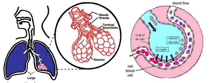
Oxygen diffuses from the alveoli to the blood in the capillaries that surround the alveoli. At the same time, carbon dioxide diffuses in the opposite direction, from capillary blood to the alveoli. At this point, the pulmonary blood is oxygen-rich, and the lungs are holding carbon dioxide. Exhalation follows, thereby ridding the body of the carbon dioxide and completing the cycle of respiration.
The taking in and expelling out of air is done by two movements:Inspiration and expiration. Inspiration: breathing in (a.k.a., inhalation)
Taking air into the body through the nose and mouth. It’s always an active process
caused by muscular contraction, mainly of the diaphragm with the help of intercostal muscles. During inhalation, the diaphragm contracts and moves downward. The rib muscles contract and cause the ribs to move outward. This causes the chest volume to increase. Because the chest volume is larger, the air pressure inside the lungs is less than the air pressure outside. This difference in air pressures causes air to be sucked into the lungs.
Expiration: breathing out (a.k.a., exhalation)Pushing air out of the body through the nose or mouth It’s typically a passive process caused by elastic recoil of the lungs and relaxation of diaphragm and intercostal muscles. When the diaphragm and rib muscles relax, the chest volume is smaller, the air pressure inside the lungs is bigger than the air pressure outside. This difference in air pressures causes air to be pushed out of the lungs. Exhalation is similar to letting the air out of a balloon.
The lungs cannot move by themselves. As mentioned above, air moves into and out of the lungs by the movement of muscles. The diaphragm and rib muscles contract and relax to move air in to and out of the lungs.
After ventilation, the second stage of breathing is the gas exchange. It takes place in the alveoli. The walls of the alveoli are very thin and are permeable to gases. The alveoli are lined with capillaries, the walls of which are also thin enough to allow gas exchange.
Oxygen diffuses from the alveoli to the blood in the capillaries that surround the alveoli. At the same time, carbon dioxide diffuses in the opposite direction, from capillary blood to the alveoli. At this point, the pulmonary blood is oxygen-rich, and the lungs are holding carbon dioxide. Exhalation follows, thereby ridding the body of the carbon dioxide and completing the cycle of respiration.
A really good finding
I am very happy about this finding. It is a page with a lot of information about different subjects. The good thing is that this page have drawing, text and the sound. You can hear the text at the same time you read it. I think is perfect to learn anything and in English.
So I must recomend this web page schoool.co.uk . In this page site you can find a good explanation of the respiratory system. You only have to click in Lesson - Breathing and Respiration and you will get several page about respiration. I suggest from page 1 to 15 to study the respiratory system and also the page 28 and 29 about smoking.
I think is also a good idea to click in Lesson - digestion to go to the digestive system pages and take a look from the page 8 to the 19. After this you will understand very much better the digestive process.
I encourage you all to use this resource. Perhaps you don't understand one word or more but as you can read the words you will be able to look up in the dictionary . I think that most of the times you will be able to guess the meaning according to the context.
Respiratory system.
Hello everybody, here we are in a new year. I hope you rested in Christmas holidays after all the work of the first quarter. I also assume that you had a good time with your family and friends. But we are in a new year and we must return to work. We have to keep improving our knowing about the human body. In the first quarter we finished with the digestive system. we studied its anatomy and fisiology. Now we continue with the nutrition function and we will study:
The respiratory system:
The main function of the respiratory system is to bring oxygen into the body and releases carbon dioxide into the atmosphere. The respiratory system is made up of the organs that take part in this process. These structures include your nose, mouth, larynx, pharynx, lungs, and diaphragm. These structures are shown in Figure

The nose and nasal cavity filters, warms, and moistens the inhaled air. The nose hairs and mucus produced by the cells that line the nose catch airborne particles and prevent them from reaching the lungs.
Behind the nasal cavity, air next passes through the pharynx, a tube that is shared with the digestive system. Both food and air pass through the pharynx. A flap of connective tissue called the epiglottis closes over the trachea when food is swallowed to prevent choking or inhaling food.
The larynx is found just below the point at which the pharynx splits into the trachea and the esophagus. Your voice comes from your larynx. Air from the lungs passes across thin membranes (vocal cords) in the larynx and produces sound.
The trachea, or wind pipe, is a long tube that leads down to the chest where it divides into the right and left bronchi in the lungs. The bronchi branch out into smaller bronchioles in each lung.
The bronchioles lead to the alveoli.
Alveoli are the little sacs at the end of the bronchioles. They look like little bunches of grapes at the end of the bronchioles, as shown in Figure. Gas exchange occurs in the alveoli, oxygen move across a membrane and into the blood and carbon dioxide move out of the blood. The alveoli are the tiny grape-like structures in the lungs and the sites of gas exchange.
The diaphragm is a sheet of muscle that extends across the bottom of the rib cage. It performs an important function in respiration. When the diaphragm contracts the chest volume gets larger and the lungs take in air. When the diaphragm relaxes, the chest volume gets smaller and air is pushed out of the lungs.
The respiratory system:
The main function of the respiratory system is to bring oxygen into the body and releases carbon dioxide into the atmosphere. The respiratory system is made up of the organs that take part in this process. These structures include your nose, mouth, larynx, pharynx, lungs, and diaphragm. These structures are shown in Figure
The nose and nasal cavity filters, warms, and moistens the inhaled air. The nose hairs and mucus produced by the cells that line the nose catch airborne particles and prevent them from reaching the lungs.
Behind the nasal cavity, air next passes through the pharynx, a tube that is shared with the digestive system. Both food and air pass through the pharynx. A flap of connective tissue called the epiglottis closes over the trachea when food is swallowed to prevent choking or inhaling food.
The larynx is found just below the point at which the pharynx splits into the trachea and the esophagus. Your voice comes from your larynx. Air from the lungs passes across thin membranes (vocal cords) in the larynx and produces sound.
The trachea, or wind pipe, is a long tube that leads down to the chest where it divides into the right and left bronchi in the lungs. The bronchi branch out into smaller bronchioles in each lung.
The bronchioles lead to the alveoli.
Alveoli are the little sacs at the end of the bronchioles. They look like little bunches of grapes at the end of the bronchioles, as shown in Figure. Gas exchange occurs in the alveoli, oxygen move across a membrane and into the blood and carbon dioxide move out of the blood. The alveoli are the tiny grape-like structures in the lungs and the sites of gas exchange.
The diaphragm is a sheet of muscle that extends across the bottom of the rib cage. It performs an important function in respiration. When the diaphragm contracts the chest volume gets larger and the lungs take in air. When the diaphragm relaxes, the chest volume gets smaller and air is pushed out of the lungs.
Suscribirse a:
Comentarios (Atom)